. 2011 Nov;79(11):4392-400.
doi: 10.1128/IAI.05478-11. Epub 2011 Aug 29.

The deubiquitinase activity of the Salmonella pathogenicity island 2 effector, SseL, prevents accumulation of cellular lipid droplets

Affiliations

The deubiquitinase activity of the Salmonella pathogenicity island 2 effector, SseL, prevents accumulation of cellular lipid droplets

Ellen T Arena et al. Infect Immun. 2011 Nov.

Abstract

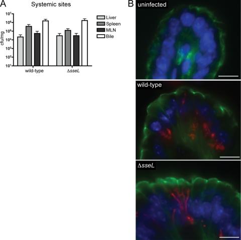
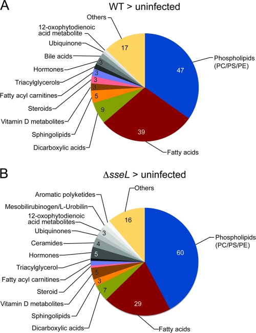
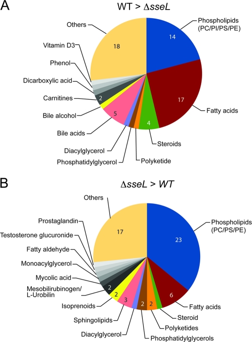
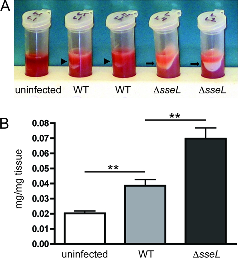
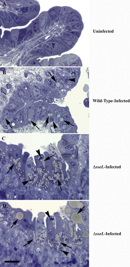
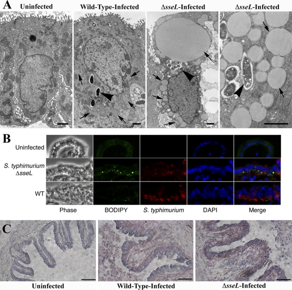
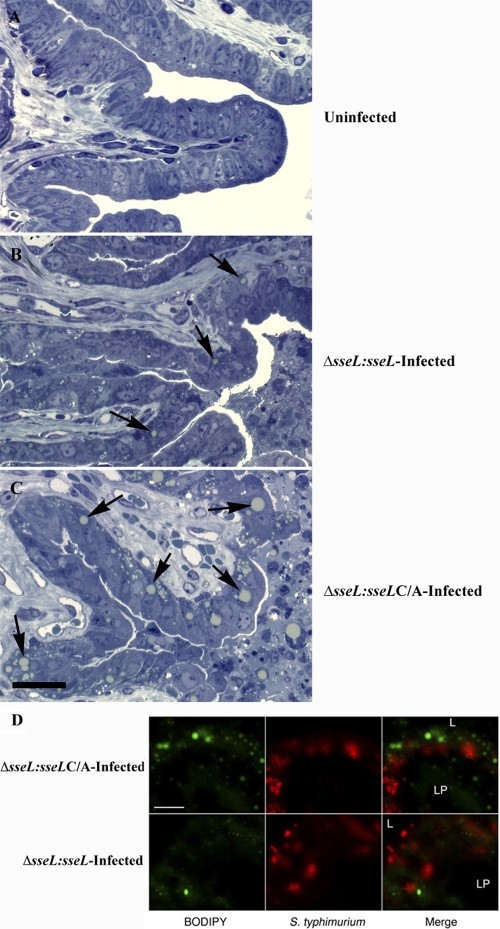
To cause disease, Salmonella enterica serovar Typhimurium requires two type III secretion systems that are encoded by Salmonella pathogenicity islands 1 and 2 (SPI-1 and -2). These secretion systems serve to deliver specialized proteins (effectors) into the host cell cytosol. While the importance of these effectors to promote colonization and replication within the host has been established, the specific roles of individual secreted effectors in the disease process are not well understood. In this study, we used an in vivo gallbladder epithelial cell infection model to study the function of the SPI-2-encoded type III effector, SseL. The deletion of the sseL gene resulted in bacterial filamentation and elongation and the unusual localization of Salmonella within infected epithelial cells. Infection with the ΔsseL strain also caused dramatic changes in host cell lipid metabolism and led to the massive accumulation of lipid droplets in infected cells. This phenotype was directly attributable to the deubiquitinase activity of SseL, as a Salmonella strain carrying a single point mutation in the catalytic cysteine also resulted in extensive lipid droplet accumulation. The excessive buildup of lipids due to the absence of a functional sseL gene also was observed in murine livers during S. Typhimurium infection. These results suggest that SseL alters host lipid metabolism in infected epithelial cells by modifying the ubiquitination patterns of cellular targets.


Figures

Fig. 1.
Fig. 1.
ΔsseL mutant strain of Salmonella is able to colonize systemic sites to wild-type levels after oral infection and is characterized by an elongated, filamentous phenotype when intracellular. (A) Bacterial counts after oral infections are presented as CFU per mg of tissue. Error bars represent standard errors from the means; all mice were sacrificed on day 5 postinfection (n=10 to 20). There are no statistical differences between wild-type and ΔsseL strain counts in the liver, spleen, mesenteric lymph nodes (MLN), or bile. Bacterial counts of bile are used as an indication of gallbladder colonization levels. (B) Immunostaining of uninfected, wild-type-infected, and ΔsseL-infected gallbladder sections collected at day 5 postinfection. Bacteria are shown in red, actin in green, and cell nuclei in blue. Scale bars indicate 10 μm.
Fig. 2.
Fig. 2.
Metabolites enriched in wild-type- or ΔsseL-infected gallbladders and uninfected gallbladders. Pie charts indicate the categories and total numbers of metabolites that are >2-fold enriched in wild-type-infected or ΔsseL-infected gallbladders after direct comparison to uninfected gallbladders. PC, phosphatidylcholine; PS, phosphatidylserine; PE, phosphatidylethanolamine.
Fig. 3.
Fig. 3.
Metabolites enriched in wild-type- or ΔsseL-infected gallbladders. Pie charts indicate the categories and total numbers of metabolites that are >2-fold enriched in wild-type-infected or ΔsseL-infected gallbladders after direct comparison to each other. “Others” includes metabolites that could not be annotated using the databases outlined in Materials and Methods. PC, phosphatidylcholine; PS, phosphatidylserine; PI, phosphatidylinositol; PE, phosphatidylethanolamine.
Fig. 4.
Fig. 4.
SseL prevents accumulation of lipids in the liver. (A) Photograph of centrifuged liver homogenates from uninfected, wild-type-infected, and ΔsseL-infected mice collected at day 5 postinfection. The lipid layer is indicated by arrowheads for the wild type and arrows for ΔsseL-infected tissues. (B) Weight of dry chloroform-methanol-soluble material (in mg) per weight of wet liver tissue (in mg) for uninfected, wild-type-infected, and ΔsseL-infected mice. Animals were infected orally, and livers were collected at day 5 postinfection; each group contained four mice. Error bars represent standard errors from the means. **, P < 0.01.
Fig. 5.
Fig. 5.
Absence of the secreted SseL effector protein results in massive accumulation of lipids within infected cells. Toluidine blue-stained gallbladder sections from an uninfected mouse (A), a wild-type-infected mouse at day 5 postinfection (B), and two representative ΔsseL mutant strain-infected mice at day 5 postinfection (C and D). Arrows indicate lipids induced upon infection, and arrowheads indicate intracellular bacteria. The scale bar indicates 10 μm.
Fig. 6.
Fig. 6.
Lipids accumulating within ΔsseL-infected gallbladder epithelial cells are in the form of lipid droplets. (A) Electron micrographs of uninfected, wild-type-infected, and ΔsseL-infected gallbladder epithelial cells at day 5 postinfection. Arrows indicate lipid droplets; arrowheads indicate intracellular bacteria. Scale bars indicate 1 μm. (B) Immunostainings of gallbladder sections collected at day 5 postinfection. Bacteria are shown in red, BODIPY in green, and cell nuclei in blue; scale bars indicate 10 μm. (C) Oil-Red-O stainings of uninfected, wild-type-infected, and ΔsseL-infected gallbladder sections collected at day 5 postinfection. Lipid droplets stain red; scale bars indicate 50 μm.
Fig. 7.
Fig. 7.
Deubiquitinase activity of SseL is essential to prevent accumulation of lipid droplets in infected cells. (A to C) Toluidine blue-stained sections of gallbladders from an uninfected mouse (A), a representative orally ΔsseL-sseL-infected mouse at day 5 postinfection (B), and a representative orally ΔsseL-sseL C/A-infected mouse at day 5 postinfection (C). Arrows indicate lipid droplets induced upon infection; the scale bar indicates 25 μm. (D) Immunostainings of the gallbladder epithelial cell layer. Tissues were collected at day 5 after oral infection. Bacteria are shown in red, and BODIPY is in green. Scale bars indicate 10 μm. L, lumen; LP, lamina propria.

References

    1. Amako Y., Sarkeshik A., Hotta H., Yates J., III, Siddiqui A. 2009. Role of oxysterol binding protein in hepatitis C virus infection. J. Virol. 83:9237–9246 - PMC - PubMed
    1. Amako Y., Syed G. H., Siddiqui A. 2011. Protein kinase D negatively regulates hepatitis C virus secretion through phosphorylation of oxysterol-binding protein and ceramide transfer protein. J. Biol. Chem. 286:11265–11274 - PMC - PubMed
    1. Antunes L. C., et al. 2011. Metabolomics reveals phospholipids as important nutrient sources during Salmonella growth in bile in vitro and in vivo. J. Bacteriol. 193:4719–4725 - PMC - PubMed
    1. Antunes L. C., et al. 2011. Impact of salmonella infection on host hormone metabolism revealed by metabolomics. Infect. Immun. 79:1759–1769 - PMC - PubMed
    1. Auweter S. D., et al. 2011. Quantitative mass spectrometry catalogues salmonella pathogenicity island-2 effectors and identifies their cognate host binding partners. J. Biol. Chem. 286:24023–24035 - PMC - PubMed

Publication types

MeSH terms

Associated data