Review
. 2011 Oct 1;3(10):a009712.
doi: 10.1101/cshperspect.a009712.

The thrombospondins

Affiliations
Review

The thrombospondins

Josephine C Adams et al. Cold Spring Harb Perspect Biol. .

Abstract

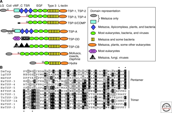
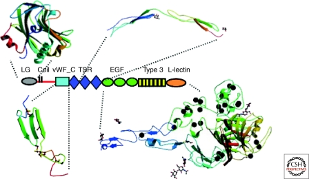
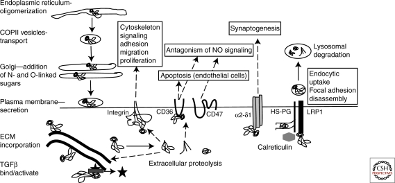
Thrombospondins are evolutionarily conserved, calcium-binding glycoproteins that undergo transient or longer-term interactions with other extracellular matrix components. They share properties with other matrix molecules, cytokines, adaptor proteins, and chaperones, modulate the organization of collagen fibrils, and bind and localize an array of growth factors or proteases. At cell surfaces, interactions with an array of receptors activate cell-dependent signaling and phenotypic outcomes. Through these dynamic, pleiotropic, and context-dependent pathways, mammalian thrombospondins contribute to wound healing and angiogenesis, vessel wall biology, connective tissue organization, and synaptogenesis. We overview the domain organization and structure of thrombospondins, key features of their evolution, and their cell biology. We discuss their roles in vivo, associations with human disease, and ongoing translational applications. In many respects, we are only beginning to appreciate the important roles of these proteins in physiology and pathology.


Figures

Figure 1.
Figure 1.
Domain architectures of thrombospondins. (A) Schematic diagram of the domain architectures of thrombospondin family members. Key: LG = laminin G-like amino-terminal domain; vWF_C = von Willebrand type C domain; TSR = thrombospondin type 1 domains; EGF = epidermal growth factor-like domains; Type 3 = thrombospondin type 3 repeats; L-lectin = L-type lectin-like domain; DD = discoidin domain; IVR = intervening region; CX2C = Cys-X2-Cys domain; CB = chitin-binding type 2 domain. Horizontal red lines indicate coiled-coil domains. Vertical black lines indicate position of cysteine residues that form intersubunit disulfide bonds. (B) Examples of the coiled-coil oligomerization domain from representative trimeric and pentameric thrombospondins. Asterisks indicate cysteines that form intersubunit disulfide bonds.
Figure 2.
Figure 2.
Structures of the domains of subgroup A thrombospondins. The crystal structures of LG (PDB 2ERF) and the second and third TSRs (PDB 1LSL) of TSP-1, and the carboxy-terminal region/signature domain of TSP-2 (PDB 1YO8) are shown. The vWF_C domain of TSP-1 is modeled on the solution structure of the vWF_C domain of collagen IIA (PDB 1U5M). Each domain is shown in a color gradient from blue at the amino terminus to red at the carboxyl terminus. The black spheres represent calcium ions. Note that the domains are not shown at the same scale.
Figure 3.
Figure 3.
Model for the evolution of thrombospondins within the metazoa. FSGD = fish-specific genome duplication. (Diagram is a development of a figure originally published in Bentley and Adams [2010]. It is reprinted, with permission, from Oxford University Press © 2010.)
Figure 4.
Figure 4.
Overview of cellular pathways and activities of mammalian TSP-1 (not to scale).

References

    1. Adams JC 2001. Thrombospondins: Multifunctional regulators of cell interactions. Annu Rev Cell Dev Biol 17: 25–51 - PubMed
    1. Adams JC 2004. Functions of the conserved thrombospondin carboxy-terminal cassette in cell-extracellular matrix interactions and signaling. Int J Biochem Cell Biol 36: 1102–1114 - PubMed
    1. Adams J, Lawler J 1993. Extracellular matrix: The thrombospondin family. Curr Biol 3: 188–190 - PubMed
    1. Adams JC, Tucker RP 2000. The thrombospondin type 1 repeat (TSR) superfamily: Diverse proteins with related roles in neuronal development. Dev Dyn 218: 280–299 - PubMed
    1. Adams JC, Clelland JD, Collett GD, Matsumura F, Yamashiro S, Zhang L 1999. Cell-matrix adhesions differentially regulate fascin phosphorylation. Mol Biol Cell 10: 4177–4190 - PMC - PubMed

Publication types

Substances

LinkOut - more resources