. 2011 Nov;31(21):4335-47.
doi: 10.1128/MCB.06075-11. Epub 2011 Aug 29.

Calpain-1 cleaves Rad21 to promote sister chromatid separation

Affiliations

Calpain-1 cleaves Rad21 to promote sister chromatid separation

Anil K Panigrahi et al. Mol Cell Biol. 2011 Nov.

Abstract

Defining the mechanisms of chromosomal cohesion and dissolution of the cohesin complex from chromatids is important for understanding the chromosomal missegregation seen in many tumor cells. Here we report the identification of a novel cohesin-resolving protease and describe its role in chromosomal segregation. Sister chromatids are held together by cohesin, a multiprotein ring-like complex comprised of Rad21, Smc1, Smc3, and SA2 (or SA1). Cohesin is known to be removed from vertebrate chromosomes by two distinct mechanisms, namely, the prophase and anaphase pathways. First, PLK1-mediated phosphorylation of SA2 in prophase leads to release of cohesin from chromosome arms, leaving behind centromeric cohesins that continue to hold the sisters together. Then, at the onset of anaphase, activated separase cleaves the centromeric cohesin Rad21, thereby opening the cohesin ring and allowing the sister chromatids to separate. We report here that the calcium-dependent cysteine endopeptidase calpain-1 is a Rad21 peptidase and normally localizes to the interphase nuclei and chromatin. Calpain-1 cleaves Rad21 at L192, in a calcium-dependent manner. We further show that Rad21 cleavage by calpain-1 promotes separation of chromosome arms, which coincides with a calcium-induced partial loss of cohesin at several chromosomal loci. Engineered cleavage of Rad21 at the calpain-cleavable site without activation of calpain-1 can lead to a loss of sister chromatid cohesion. Collectively, our work reveals a novel function of calpain-1 and describes an additional pathway for sister chromatid separation in humans.


Figures

Fig. 1.
Fig. 1.
Rad21 is a substrate of calpain-1. (A) Myc.Rad21, synthesized in vitro using wheat germ extract, was incubated with 0, 0.2, 0.5, 1.25, and 1.25 mU of purified calpain-1 (lanes 1 to 5, respectively); ALLN was added to the last reaction mix at a 5 μM final concentration (lane 5). Full-length (FL) and cleaved (Cl.) Rad21 bands are indicated by arrows. (B) Molt4 cells were treated with CaCl2 (2 mM final concentration) for 5 h (lane 2) or additionally pretreated with 5 μM ALLN (lane 3). WCE was probed with the indicated antibodies. (C) (Top) Schematic of hRad21 and relative location of the calpain-1 cleavage site (not drawn to scale). The inverted arrow indicates the fissile bond. (Bottom) Phylogenetic sequence comparison of Rad21 sequences relative to the sequence of the human protein (amino acids [aa] 168 to 202). Numbers on the left of gels indicate molecular masses in kDa.
Fig. 2.
Fig. 2.
Cleavage of Rad21 by calpain-1. (A) Amino acid sequences flanking the calpain-cleavable site (relative to aa 184 to 202) in WT and mutant Rad21. Replacement residues and the TEV cleavage sequence are highlighted in italics and bold, respectively. (B) WT and mutant Myc.Rad21 proteins were synthesized in vitro and incubated with 1 mU calpain-1. A nonspecific band is marked with an asterisk. (C) WT and tev mutant Myc.Rad21 proteins were incubated with or without 0.5 U of TEV protease (TEV). (D) HEK293T cells were transfected with empty vector (EV) or a plasmid carrying WT or del mutant Myc.Rad21. Endogenous Rad21 was knocked down by siRNA duplexes targeting the hRad21 3′-UTR. At 2 days posttransfection, cells were treated with 10 mM CaCl2 for 6 h. (E) HEK293T cells were transfected with control or calpain-1-specific siRNA (duplex A) and treated with 10 mM CaCl2 for 6 h at 3 days posttransfection.
Fig. 3.
Fig. 3.
Calcium-induced Rad21 cleavage is not associated with apoptosis or necrosis. (A) Molt4 cells were pretreated with etoposide (Etop; 20 μM final concentration) or ALLN (5 μM final concentration) for 30 min before addition of CaCl2 (2 mM final concentration). Cells were harvested after 6 h, and WCE was prepared. Note that the calcium-induced cleavage product of Rad21 (Cl. Rad21) was different from the apoptotic cleavage products (Apopt. Cl. Rad21). Nonspecific bands are marked with asterisks. (B) Molt4 cells were treated with 2 mM CaCl2 or 5 μM etoposide for 28 h, stained with annexin V-FITC and propidium iodide, and observed using light as well as fluorescence microscopy. The graph on the right shows the number of apoptotic cells as a percentage of the total number of cells.
Fig. 4.
Fig. 4.
Calpain-1 localizes to chromatin and cleaves chromatin-bound Rad21. (A) Scheme of cell fractionation. Cells were pretreated with ALLN (final concentration, 5 μM) for 30 min before the addition of CaCl2 (2 mM) for 10 h. (B) Nuclear extract (Nuc. Extr.) and chromatin fractions were resolved and probed with the indicated antibodies. (C) Fractionation of Rad21 cleavage upon calcium treatment. Jurkat cells, treated in the presence or absence of 2 mM CaCl2 for the indicated times (hours), were fractionated into cytosolic extract (CE), nuclear extract (NE), and chromatin (Chr) fractions. Proteins of equal volumetric proportions were analyzed by immunoblotting. Full-length (FL) and cleaved (Cl.) Rad21 proteins are indicated. α-Tubulin, lamin B, and histone H3 serve as markers for cell fractionation. (D) Immunofluorescence staining of HeLa cells by use of the indicated antibodies before (top) and after (bottom) extraction with Triton X-100.
Fig. 5.
Fig. 5.
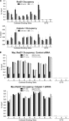
Calpain-1 activation affects Rad21 occupancy. (A) Chromatin occupancy of Rad21 and calpain-1 in Jurkat cells. Rad21 and calpain-1 occupancy is shown for nine cohesin binding sites and three cohesin-free sites (CFS). (B) Chromatin occupancy of Myc-tagged WT and del mutant Rad21 in HEK293T cells transfected with control (top) or calpain-1 (duplex A) (bottom) siRNA. Average threshold cycle (CT) values for each primer pair for three independent biological replicates (each run in triplicate) were analyzed. Error bars represent standard errors of the means.
Fig. 6.
Fig. 6.
Calpain-1 affects sister chromatid separation. (A) Four major types of metaphase were taken into account. (B) Jurkat cells were treated with 2 mM CaCl2 for 2 and 4 days and then subjected to protein analysis (top) and metaphase spread analysis (bottom) along with untreated controls. Various metaphase types were scored. (C) HEK293T cells were transfected with control (Ctr) siRNA duplexes and with siRNAs targeting Rad21, separase (Sep), and calpain-1 (Calp1; duplex A). Cells were harvested at 72 h posttransfection. Protein (top) and metaphase spread (bottom) analyses are shown. (D) HeLa cells were transfected with control and calpain-1 (duplex A) siRNAs and were treated with 10 mM CaCl2 at 2 days posttransfection. Cells were analyzed for protein profiles by immunoblotting (left) and for DNA content by FACS (right). (E) HEK293T cells were transfected with either one of the three independent calpain-1 siRNAs (duplexes A, B, and C) or with control siRNA and were treated with CaCl2 at 48 h posttransfection. Protein profiles (top) and metaphase chromosome analysis (bottom) are shown. Nonspecific bands are marked with asterisks.
Fig. 7.
Fig. 7.
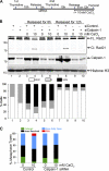
(A) Schematic of synchronization and release of HeLa cells following double thymidine block. (B) HeLa cells were synchronized by double thymidine arrest, transfected with calpain-1 siRNA duplex A after the first thymidine block, and finally released for 8 h with or without an additional 10 mM CaCl2. Protein profiles (for whole-cell lysates) (top) and percentages of cells as analyzed by FACS (bottom) are shown. Histone H3 is shown as a loading control. Lanes 1 and 2 represent cells arrested at the G1/S transition. (C) Metaphase spread analysis of cells harvested 8 h after release from the double thymidine block. Cells from a single 10-cm plate were split equally and processed for protein, FACS, and metaphase spread analyses.
Fig. 8.
Fig. 8.
HEK293T cells were first transfected with a plasmid for WT Myc.Rad21 (WT) or Myc.Rad21tev (tev) along with pCS2MT-TEV (pTEV), as indicated. Twenty-four hours later, cells were transfected with siRNA duplexes targeting the 3′-UTR of Rad21. Cells were grown for a further 48 h before harvesting. Protein profile (A) and metaphase spread (B) analyses are shown.
Fig. 9.
Fig. 9.
Model for calpain-1-mediated Rad21 cleavage and sister chromatid separation. Cleavage of either one or both Rad21 molecules (represented by white incisions) in the cohesin handcuff by calpain-1 (CAPN1) can equally lead to a loss of sister chromatid cohesion and can also relieve cohesin-mediated catenation of sisters. Cohesin cleavage by calpain-1 may function as an additional mechanism to separate the sisters, aiding the Plk1-mediated prophase pathway.

Similar articles

Cited by

References

    1. Beckouet F., et al. 2010. An Smc3 acetylation cycle is essential for establishment of sister chromatid cohesion. Mol. Cell 39:689–699 - PMC - PubMed
    1. Birkenbihl R. P., Subramani S. 1992. Cloning and characterization of rad21 an essential gene of Schizosaccharomyces pombe involved in DNA double-strand-break repair. Nucleic Acids Res. 20:6605–6611 - PMC - PubMed
    1. Bootman M. D., Fearnley C., Smyrnias I., MacDonald F., Roderick H. L. 2009. An update on nuclear calcium signalling. J. Cell Sci. 122:2337–2350 - PubMed
    1. Bootman M. D., Young K. W., Young J. M., Moreton R. B., Berridge M. J. 1996. Extracellular calcium concentration controls the frequency of intracellular calcium spiking independently of inositol 1,4,5-trisphosphate production in HeLa cells. Biochem. J. 314:347–354 - PMC - PubMed
    1. Borges V., et al. 2010. Hos1 deacetylates Smc3 to close the cohesin acetylation cycle. Mol. Cell 39:677–688 - PubMed

Publication types

MeSH terms

LinkOut - more resources