

Disruption of K(2P)6.1 produces vascular dysfunction and hypertension in mice
- PMID: 21876070
- PMCID: PMC3205080
- DOI: 10.1161/HYPERTENSIONAHA.111.175349
Disruption of K(2P)6.1 produces vascular dysfunction and hypertension in mice
Abstract
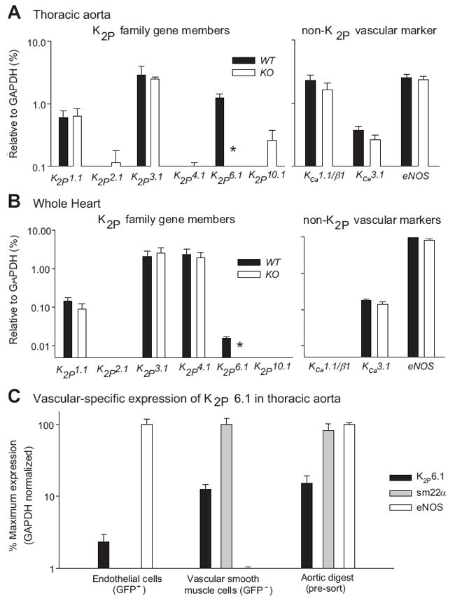
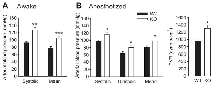
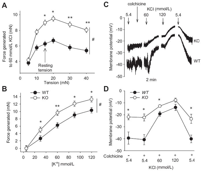
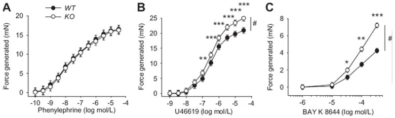
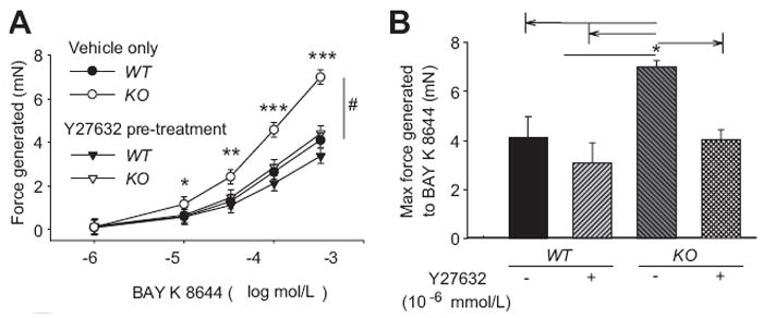
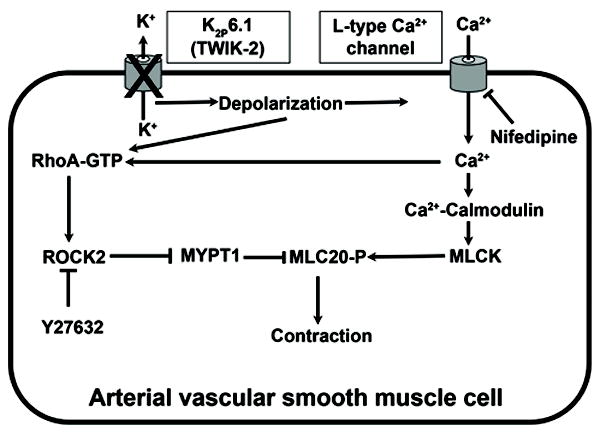
K(2P)6.1, a member of the 2-pore domain K channel family, is highly expressed in the vascular system; however, its function is unknown. We tested the following hypotheses. K(2P)6.1 regulates the following: (1) systemic blood pressure; (2) the contractile state of arteries; (3) vascular smooth muscle cell migration; (4) proliferation; and/or (5) volume regulation. Mice lacking K(2P)6.1 (KO) were generated by deleting exon 1 of Kcnk6. Mean arterial blood pressure in both anesthetized and awake KO mice was increased by 17±2 and 26±3 mm Hg, respectively (P<0.05). The resting membrane potential in freshly dispersed vascular smooth muscle cells was depolarized by 17±2 mV in the KO compared with wild-type littermates (P<0.05). The contractile responses to KCl (P<0.05) and BAY K 8644 (P<0.01), an activator of L-type calcium channels, were enhanced in isolated segments of aorta from KO mice. However, there was no difference in the current density of L-type calcium channels. Responses to U46619, an agent that activates rho kinase, showed an enhanced contraction in aorta from KO mice (P<0.001). The BAY K 8644-mediated increase in contraction was decreased to wild-type levels when treated with Y27632, a rho kinase inhibitor, (P<0.05). K(2P)6.1 does not appear to be involved with migration, proliferation, or volume regulation in cultured vascular smooth muscle cells. We conclude that K(2P)6.1 deficiency induces vascular dysfunction and hypertension through a mechanism that may involve smooth muscle cell depolarization and enhanced rho kinase activity.
Conflict of interest statement
Conflict(s) of interest/Disclosure(s) Statement None
Figures
Comment in
-
Two-pore domain K⁺ channels: evidence for TWIK-2 in blood pressure regulation.Hypertension. 2011 Oct;58(4):539-41. doi: 10.1161/HYPERTENSIONAHA.111.179390. Epub 2011 Aug 29. Hypertension. 2011. PMID: 21876073 No abstract available.
References
-
- Hille B. Ionic Channels of Excitable Membranes. 3. Sunderland, Mass: 2001.
-
- Bryan RM, Jr, Joseph BK, Lloyd E, Rusch NJ. Starring TREK-1: the next generation of vascular K+ channels. Circ Res. 2007;101:119–121. - PubMed
-
- Goldstein SA, Bayliss DA, Kim D, Lesage F, Plant LD, Rajan S. International Union of Pharmacology. LV. Nomenclature and molecular relationships of two-P potassium channels. Pharmacol Rev. 2005;57:527–540. - PubMed
-
- Chavez RA, Gray AT, Zhao BB, Kindler CH, Mazurek MJ, Mehta Y, Forsayeth JR, Yost CS. TWIK-2, a new weak inward rectifying member of the tandem pore domain potassium channel family. J Biol Chem. 1999;274:7887–7892. - PubMed
-
- Patel AJ, Maingret F, Magnone V, Fosset M, Lazdunski M, Honore E. TWIK-2, an inactivating 2P domain K+ channel. J Biol Chem. 2000;275:28722–28730. - PubMed
Publication types
MeSH terms
Substances
Grants and funding
LinkOut - more resources
Full Text Sources
Medical
Molecular Biology Databases
Research Materials
