FGF signaling is required for lens regeneration in Xenopus laevis
- PMID: 21876116
- PMCID: PMC3442785
- DOI: 10.1086/BBLv221n1p137
FGF signaling is required for lens regeneration in Xenopus laevis
Abstract
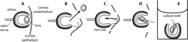
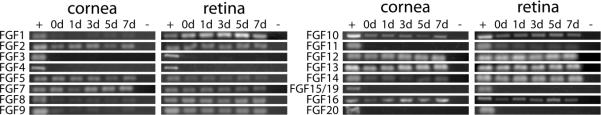
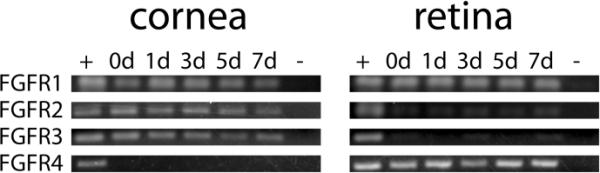
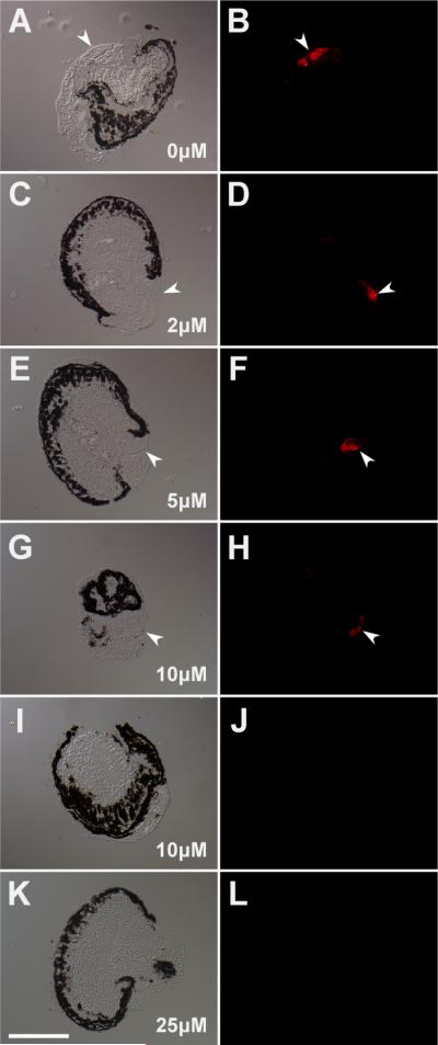
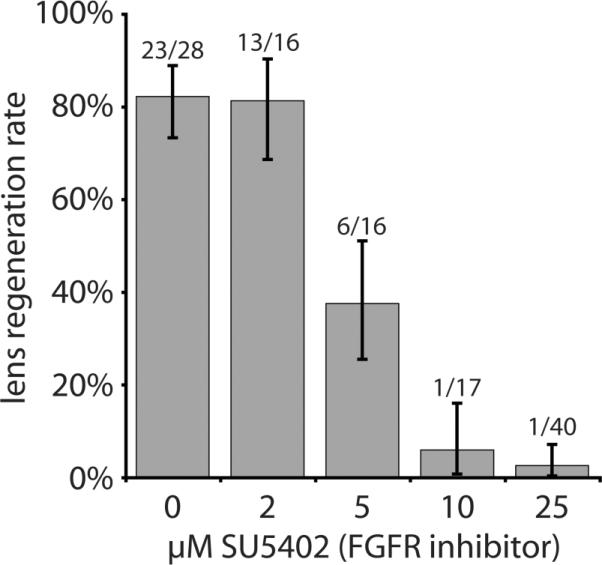
In species of the frog genus Xenopus, lens regeneration occurs through a process of transdifferentiation, in which cornea epithelial cells presumably undergo dedifferentiation and subsequently redifferentiate to form a new lens. Experimental studies have shown that the retina provides the key signal required to trigger this process once the original lens is removed. A previous study showed that addition of an exogenous fibroblast growth factor (i.e., FGF1 protein) could initiate transdifferentiation of cornea epithelial cells in culture. To determine the role of FGF signaling in X. laevis lens regeneration, we have examined the presence of specific FGFs and their receptors (FGFRs) during this process and evaluated the necessity of FGFR signaling. Reverse transcriptase-polymerase chain reaction analyses reveal that a number of FGF family members are expressed in cornea epithelium and retinal tissues both before and during the process of lens regeneration. Of these, FGF1, FGF8, and FGF9 are expressed principally in retinal tissue and not in the cornea epithelium. Hence, these ligands could represent key signaling factors originating from the retina that trigger regeneration. The results of experiments using an in vitro eye culture system and an FGFR inhibitor (SU5402) suggest that FGFR signaling is required for lens regeneration in Xenopus.
Figures
Similar articles
-
Retinoic acid regulation by CYP26 in vertebrate lens regeneration.Dev Biol. 2014 Feb 15;386(2):291-301. doi: 10.1016/j.ydbio.2013.12.036. Epub 2013 Dec 30. Dev Biol. 2014. PMID: 24384390 Free PMC article.
-
Expression of pluripotency factors in larval epithelia of the frog Xenopus: evidence for the presence of cornea epithelial stem cells.Dev Biol. 2013 Feb 15;374(2):281-94. doi: 10.1016/j.ydbio.2012.12.005. Epub 2012 Dec 26. Dev Biol. 2013. PMID: 23274420 Free PMC article.
-
Cornea-lens transdifferentiation in the anuran, Xenopus tropicalis.Dev Genes Evol. 2001 Sep;211(8-9):377-87. doi: 10.1007/s004270100163. Dev Genes Evol. 2001. PMID: 11685571
-
An essential role for FGF receptor signaling in lens development.Semin Cell Dev Biol. 2006 Dec;17(6):726-40. doi: 10.1016/j.semcdb.2006.10.002. Epub 2006 Oct 27. Semin Cell Dev Biol. 2006. PMID: 17116415 Free PMC article. Review.
-
Retina and lens regeneration in anuran amphibians.Semin Cell Dev Biol. 2009 Jul;20(5):528-34. doi: 10.1016/j.semcdb.2008.11.015. Epub 2008 Nov 27. Semin Cell Dev Biol. 2009. PMID: 19095070 Review.
Cited by
-
Myogenic-specific ablation of Fgfr1 impairs FGF2-mediated proliferation of satellite cells at the myofiber niche but does not abolish the capacity for muscle regeneration.Front Aging Neurosci. 2015 May 28;7:85. doi: 10.3389/fnagi.2015.00085. eCollection 2015. Front Aging Neurosci. 2015. PMID: 26074812 Free PMC article.
-
Pigment Epithelia of the Eye: Cell-Type Conversion in Regeneration and Disease.Life (Basel). 2022 Mar 6;12(3):382. doi: 10.3390/life12030382. Life (Basel). 2022. PMID: 35330132 Free PMC article. Review.
-
Diverse Evolutionary Origins and Mechanisms of Lens Regeneration.Mol Biol Evol. 2018 Jul 1;35(7):1563-1575. doi: 10.1093/molbev/msy045. Mol Biol Evol. 2018. PMID: 29579253 Free PMC article. Review.
-
Lens regeneration from the cornea requires suppression of Wnt/β-catenin signaling.Exp Eye Res. 2016 Apr;145:206-215. doi: 10.1016/j.exer.2016.01.003. Epub 2016 Jan 8. Exp Eye Res. 2016. PMID: 26778749 Free PMC article.
-
Epigenetic Modifications in the Retinal Pigment Epithelium of the Eye During RPE-Related Regeneration or Retinal Diseases in Vertebrates.Biomedicines. 2025 Jun 25;13(7):1552. doi: 10.3390/biomedicines13071552. Biomedicines. 2025. PMID: 40722628 Free PMC article. Review.
References
-
- Arresta E, Bernardini S, Gargioli C, Filoni S, Cannata SM. Lens-forming competence in the epidermis of Xenopus laevis during development. J Exp Zool A Comp Exp Biol. 2005;303A:1–12. - PubMed
-
- Baird A, Esch F, Mormède P, Ueno N, Ling N, Böhlen P, Ying SY, Wehrenberg WB, Guillemin R. Molecular characterization of fibroblast growth factor: distribution and biological activities in various tissues. Recent Prog.Horm.Res. 1986;42:143–205. - PubMed
-
- Bosco L, Filoni S, Cannata S. Relationships between Eye Factors and Lens-forming Transformations in the Cornea and Pericorneal Epidermis of Larval Xenopus laevis. J. Exp. Zool. 1979;209:261–282. - PubMed
-
- Bosco L, Valle C, Willems D. In Vivo and In Vitro Experimental Analysis of Lens Regeneration in Larval Xenopus laevis. Dev.Growth Differ. 1993;35:257–270. - PubMed
Publication types
MeSH terms
Substances
Grants and funding
LinkOut - more resources
Full Text Sources