

Quantitative lipid composition of cell envelopes of Corynebacterium glutamicum elucidated through reverse micelle extraction
- PMID: 21876124
- PMCID: PMC3174599
- DOI: 10.1073/pnas.1112572108
Quantitative lipid composition of cell envelopes of Corynebacterium glutamicum elucidated through reverse micelle extraction
Abstract
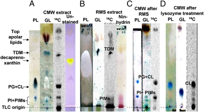
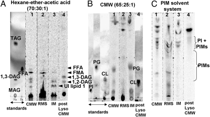
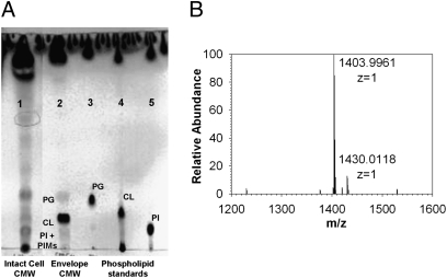
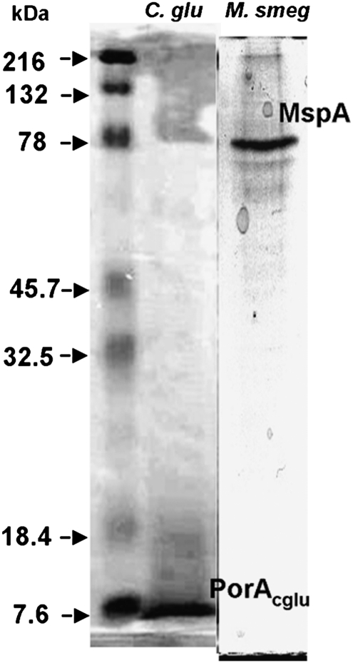
Cells of the Corynebacterium-Nocardia-Mycobacterium group of bacteria are surrounded by an outer membrane (OM) containing mycolic acids that are covalently linked to the underlying arabinogalactan-peptidoglycan complex. This OM presumably acts as a permeability barrier that imparts high levels of intrinsic drug resistance to some members of this group, such as Mycobacterium tuberculosis, and its component lipids have been studied intensively in a qualitative manner over the years. However, the quantitative lipid composition of this membrane has remained obscure, mainly because of difficulties in isolating it without contamination from the inner cytoplasmic membrane. Here we use the extraction, with reverse surfactant micelles, of intact cells of Corynebacterium glutamicum and show that this method extracts the free OM lipids quantitatively with no contamination from lipids of the cytoplasmic membrane, such as phosphatidylglycerol. Although only small amounts of corynomycolate were esterified to arabinogalactan, a large amount of cardiolipin was present in a nonextractable form, tightly associated, possibly covalently, with the peptidoglycan-arabinogalactan complex. Furthermore, we show that the OM contains just enough lipid hydrocarbons to produce a bilayer covering the cell surface, with its inner leaflet composed mainly of the aforementioned nonextractable cardiolipin and its outer leaflet composed of trehalose dimycolates, phosphatidylinositol mannosides, and highly apolar lipids, similar to the Minnikin model of 1982. The reverse micelle extraction method is also useful for extracting proteins associated with the OM, such as porins.
Conflict of interest statement
The authors declare no conflict of interest.
Figures
References
-
- Minnikin D. In: The Biology of Mycobacteria. Ratledge C, Stanford J, editors. Vol 1. London: Academic; 1982. pp. 94–184.
-
- Wheeler PR, Ratledge C. Metabolism in Mycobacterium leprae, M. tuberculosis and other pathogenic mycobacteria. Br Med Bull. 1988;44:547–561. - PubMed
-
- McNeil MR, Brennan PJ. Structure, function and biogenesis of the cell envelope of mycobacteria in relation to bacterial physiology, pathogenesis and drug resistance: Some thoughts and possibilities arising from recent structural information. Res Microbiol. 1991;142:451–463. - PubMed
-
- Nikaido H, Kim S-H, Rosenberg EY. Physical organization of lipids in the cell wall of Mycobacterium chelonae. Mol Microbiol. 1993;8:1025–1030. - PubMed
Publication types
MeSH terms
Substances
Grants and funding
LinkOut - more resources
Full Text Sources
Molecular Biology Databases
