. 2011 Sep 13;108(37):15174-8.
doi: 10.1073/pnas.1106341108. Epub 2011 Aug 29.

Correlation of in situ mechanosensitive responses of the Moraxella catarrhalis adhesin UspA1 with fibronectin and receptor CEACAM1 binding

Affiliations

Correlation of in situ mechanosensitive responses of the Moraxella catarrhalis adhesin UspA1 with fibronectin and receptor CEACAM1 binding

Christopher Agnew et al. Proc Natl Acad Sci U S A. .

Abstract

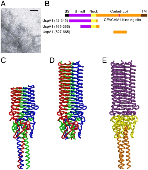
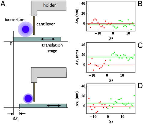
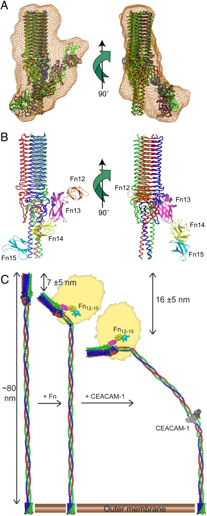
Bacterial cell surfaces are commonly decorated with a layer formed from multiple copies of adhesin proteins whose binding interactions initiate colonization and infection processes. In this study, we investigate the physical deformability of the UspA1 adhesin protein from Moraxella catarrhalis, a causative agent of middle-ear infections in humans. UspA1 binds a range of extracellular proteins including fibronectin, and the epithelial cellular receptor carcinoembryonic antigen-related cell adhesion molecule 1 (CEACAM1). Electron microscopy indicates that unliganded UspA1 is densely packed at, and extends about 800 Å from, the Moraxella surface. Using a modified atomic force microscope, we show that the adhesive properties and thickness of the UspA1 layer at the cell surface varies on addition of either fibronectin or CEACAM1. This in situ analysis is then correlated with the molecular structure of UspA1. To provide an overall model for UspA1, we have determined crystal structures for two N-terminal fragments which are then combined with a previous structure of the CEACAM1-binding site. We show that the UspA1-fibronectin complex is formed between UspA1 head region and the 13th type-III domain of fibronectin and, using X-ray scattering, that the complex involves an angular association between these two proteins. In combination with a previous study, which showed that the CEACAM1-UspA1 complex is distinctively bent in solution, we correlate these observations on isolated fragments of UspA1 with its in situ response on the cell surface. This study therefore provides a rare direct demonstration of protein conformational change at the cell surface.


Conflict of interest statement

The authors declare no conflict of interest.

Figures

Fig. 1.
Fig. 1.
Moraxella cell surface, domain organization, and head region structure of UspA1. (A) Electron micrograph showing extended UspA1 molecules at the Mx surface. (Scale bar: 50 nm.) (B) Schematic showing full length UspA1 comprises five regions, each colored separately and labeled. Constructs for which crystal structures are determined are also shown. TM, transmembrane. (CE) Ribbon representations of the crystal structures of (C) UspA1(165–366), (D) UspA1(42–345), and (E) composite model of UspA1(42–366). In C and D, each chain is colored separately; E is colored by domain as per B.
Fig. 2.
Fig. 2.
Atomic force microscopy analysis of Mx bacteria. (A) A sinusoidal sideways movement is imparted to the sample stage in order to gently push a single adsorbed bacterium (blue sphere; pale-blue outer layer represents surface adhesins) against the cantilever. The change in the sample stage position (Δx1) required to achieve contact is measured upon addition of either ligand (CEACAM1 or Fn) or buffer/control protein. The graphs on the right show representative data measured for the change in the contact point position before (in red) and after (in green) the addition of (B) control, (C) CEACAM1, and (D) FNIII12,13,14. The red and green lines represent the mean value of the red and green points, respectively.
Fig. 3.
Fig. 3.
SAXS analysis of UspA1(42–366)-FnIII12–15 complex. (A) Molecular envelope (orange) defined by ab initio bead modeling, with five independent rigid-body reconstructions shown as Cα ribbons, with each reconstruction displayed in a different color. (B) Representative rigid-body reconstruction of UspA1–Fn complex, with UspA1(165–366) colored as in Fig. 1C and FnIII12–15 as a Cα cartoon colored by domain and labeled. Heparin-binding residues are shown as spheres. (C) Overall model for UspA1(154–869) (colored as in Fig. 1C) and deformation associated with sequentially binding Fn (colored as in B) and CEACAM-1 (in gray). The remainder of the Fn molecule is symbolized as a rough yellow ellipsoid (not to scale). The distances and uncertainties shown are from the LMFM analysis and represent the decrease in overall UspA1 length resulting from binding of each ligand. The bending angle on binding CEACAM1 required to achieve this change has been calculated from the linear dimensions of the combined crystal structures and is estimated to be approximately 45 ± 15°, which is within the range of movement observed in a prior molecular dynamics simulation (7).

Similar articles

Cited by

References

    1. Karalus R, Campagnari A. Moraxella catarrhalis: A review of an important human mucosal pathogen. Microbes Infect. 2000;2:547–559. - PubMed
    1. Hultgren SJ, et al. Pilus and nonpilus bacterial adhesins: Assembly and function in cell recognition. Cell. 1993;73:887–901. - PubMed
    1. Hoiczyk E, Roggenkamp A, Reichenbecher M, Lupas A, Heesemann J. Structure and sequence analysis of Yersinia YadA and Moraxella UspAs reveal a novel class of adhesins. EMBO J. 2000;19:5989–5999. - PMC - PubMed
    1. Szczesny P, et al. Structure of the head of the Bartonella adhesin BadA. PLoS Pathog. 2008;4:e1000119. - PMC - PubMed
    1. Wells TJ, Tree JJ, Ulett GC, Schembri MA. Autotransporter proteins: Novel targets at the bacterial cell surface. FEMS Microbiol Lett. 2007;274:163–172. - PubMed

Publication types

MeSH terms

LinkOut - more resources