. 2011 Sep 13;108(37):15196-200.
doi: 10.1073/pnas.1107819108. Epub 2011 Aug 29.

Interaction of complexes I, III, and IV within the bovine respirasome by single particle cryoelectron tomography

Affiliations

Interaction of complexes I, III, and IV within the bovine respirasome by single particle cryoelectron tomography

Natalya V Dudkina et al. Proc Natl Acad Sci U S A. .

Abstract

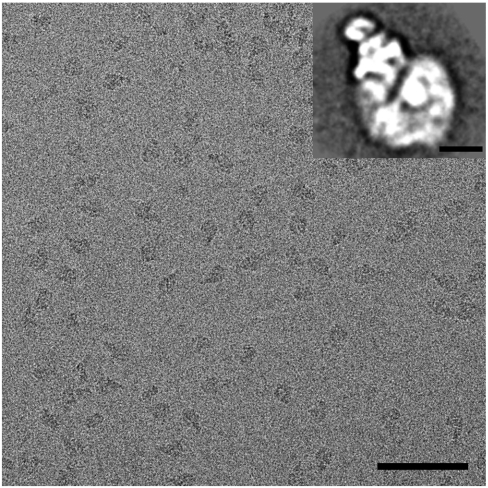
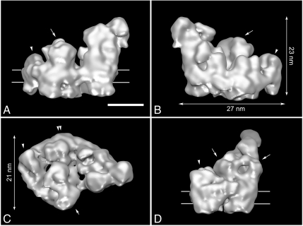
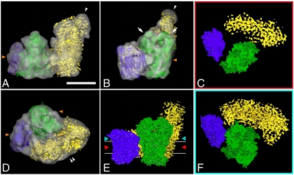
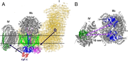
The respirasome is a multisubunit supercomplex of the respiratory chain in mitochondria. Here we report the 3D reconstruction of the bovine heart respirasome, composed of dimeric complex III and single copies of complex I and IV, at about 2.2-nm resolution, determined by cryoelectron tomography and subvolume averaging. Fitting of X-ray structures of single complexes I, III(2), and IV with high fidelity allows interpretation of the model at the level of secondary structures and shows how the individual complexes interact within the respirasome. Surprisingly, the distance between cytochrome c binding sites of complexes III(2) and IV is about 10 nm. Modeling indicates a loose interaction between the three complexes and provides evidence that lipids are gluing them at the interfaces.


Conflict of interest statement

The authors declare no conflict of interest.

Figures

Fig. 1.
Fig. 1.
Cryoelectron micrograph of isolated bovine respirasomes. (Scale bar: 100 nm.) (Inset) Two-dimensional projection map of the respirasome in a top view position from single particle image analysis. (Scale bar: 10 nm.)
Fig. 2.
Fig. 2.
Cryo-EM map of I + III2 + IV supercomplex: (A and B) seen from aside, (C) seen from the matrix side; arrow points to complex III2, arrowhead to complex IV, and double arrowhead to the curved membrane arm of complex I, respectively. (D) Seen from aside from the tip. Contour level is 0.1. Horizontal lines on A and D indicate the position of the membrane. (Scale bar: 10 nm.)
Fig. 3.
Fig. 3.
Fitting of the high- and medium-resolution structures of complexes I, III2, and IV to the 3D cryo-EM map of I + III2 + IV supercomplex: (A) side view, arrowhead points to flavoproteins; (B) side view from the membrane, arrows point to core I and II subunits of complex III2, arrowhead to flavoproteins; (C) section through the space-filling model of respirasome on the level of membrane, demonstrating gaps between complexes within the supercomplex; (D) top view from the intermembrane space, double arrowhead points to the bend of complex I in membrane; (E) space-filling model of respirasome seen from the membrane, red and light-blue arrowheads show the level of sections in C and F; (F) section through the space-filling model of respirasome on the level of matrix. In green, X-ray structure of the bovine dimeric complex III; in purple, X-ray structure of bovine monomeric complex IV; in yellow, the density map of complex I from Yarrowia lipolytica. Horizontal lines on E indicate the position of the membrane. Orange arrowheads on A, B, and D point to the position of detergent micelles. (Scale bar: 10 nm.)
Fig. 4.
Fig. 4.
Scheme of electron flow within the respirasome mediated by cytochrome c and ubiquinol. (A) Side view of the I + III2 + IV supercomplex. Black arrows point how the ubiquinol and the cytochrome c move between complexes I, III2, and IV. (B) Top view of the respirasome fragment consisting of complexes III2 and IV, seen from the intermembrane space. The distance between cytochrome c binding sites of complexes III2 and IV is 10 nm. In yellow, the density map of complex I from Yarrowia lipolytica, marked as I; in gray, X-ray structures of bovine complexes III2 and IV, marked as III2 and IV correspondingly. Q, ubiquinol; cyt c, cytochrome c. Colors: red, cytochrome c; dark green, subunit II of complex IV; blue, cytochrome c1 subunit of complex III2; green, cytochrome b of complex III2; magenta, “Rieske” protein of complex III2. Horizontal lines on A indicate the position of the membrane.

References

    1. Schägger H, Pfeiffer K. Supercomplexes in the respiratory chains of yeast and mammalian mitochondria. EMBO J. 2000;19:1777–1783. - PMC - PubMed
    1. Dudkina NV, Eubel H, Keegstra W, Boekema EJ, Braun HP. Structure of a mitochondrial supercomplex formed by respiratory-chain complexes I and III. Proc Natl Acad Sci USA. 2005;102:3225–3229. - PMC - PubMed
    1. Minauro-Sanmiguel F, Wilkens S, Garcia JJ. Structure of dimeric mitochondrial ATP synthase: Novel F0 bridging features and the structural basis of mitochondrial cristae biogenesis. Proc Natl Acad Sci USA. 2005;102:12356–12358. - PMC - PubMed
    1. Dudkina NV, Heinemeyer J, Keegstra W, Boekema EJ, Braun HP. Structure of dimeric ATP synthase from mitochondria: An angular association of monomers induces the strong curvature of the inner membrane. FEBS Lett. 2005;579:5769–5772. - PubMed
    1. Dudkina NV, Kouřil R, Peters K, Braun HP, Boekema EJ. Structure and function of mitochondrial supercomplexes. Biochim Biophys Acta. 2010;1797:664–670. - PubMed

Publication types

MeSH terms

Substances

LinkOut - more resources