. 2011 Sep 13;108(37):E709-17.
doi: 10.1073/pnas.1101655108. Epub 2011 Aug 29.

Global discovery of small RNAs in Yersinia pseudotuberculosis identifies Yersinia-specific small, noncoding RNAs required for virulence

Affiliations

Global discovery of small RNAs in Yersinia pseudotuberculosis identifies Yersinia-specific small, noncoding RNAs required for virulence

Jovanka T Koo et al. Proc Natl Acad Sci U S A. .

Abstract

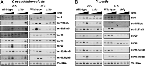
A major class of bacterial small, noncoding RNAs (sRNAs) acts by base-pairing with mRNAs to alter the translation from and/or stability of the transcript. Our laboratory has shown that Hfq, the chaperone that mediates the interaction of many sRNAs with their targets, is required for the virulence of the enteropathogen Yersinia pseudotuberculosis. This finding suggests that sRNAs play a critical role in the regulation of virulence in this pathogen, but these sRNAs are not known. Using a deep sequencing approach, we identified the global set of sRNAs expressed in vitro by Y. pseudotuberculosis. Sequencing of RNA libraries from bacteria grown at 26 °C and 37 °C resulted in the identification of 150 unannotated sRNAs. The majority of these sRNAs are Yersinia specific, without orthologs in either Escherichia coli or Salmonella typhimurium. Six sRNAs are Y. pseudotuberculosis specific and are absent from the genome of the closely related species Yersinia pestis. We found that the expression of many sRNAs conserved between Y. pseudotuberculosis and Y. pestis differs in both timing and dependence on Hfq, suggesting evolutionary changes in posttranscriptional regulation between these species. Deletion of multiple sRNAs in Y. pseudotuberculosis leads to attenuation of the pathogen in a mouse model of yersiniosis, as does the inactivation in Y. pestis of a conserved, Yersinia-specific sRNA in a mouse model of pneumonic plague. Finally, we determined the regulon controlled by one of these sRNAs, revealing potential virulence determinants in Y. pseudotuberculosis that are regulated in a posttranscriptional manner.


Conflict of interest statement

The authors declare no conflict of interest.

Figures

Fig. 1.
Fig. 1.
Experimental identification of annotated sRNAs in Y. pseudotuberculosis. (A) All previously annotated Y. pseudotuberculosis sRNAs were identified by Illumina-Solexa sequencing. Percent of total reads for annotated RNAs identified at 37 °C shows a flux in sRNA levels over the course of bacterial growth. (B) The expression levels of the sRNA spf (Spot 42) as determined by deep sequencing reads (red bars in A) correlate with the levels of the sRNA as measured by Northern blot analysis.
Fig. 2.
Fig. 2.
Yersinia small RNAs (Ysrs). The sequences of sRNAs identified by deep sequencing in Y. pseudotuberculosis strain IP32953 were compared with the genomes of Y. pestis strain CO92, Y. enterocolitica 8081, E. coli strain K12 (substrain MG1655), and Salmonella enterica serovar Typhimurium strain LT2 by BlastN analysis. Ysrs present in the genome and identical in sequence are shown in green, and Ysrs present in the genome with sequence differences are shown in yellow. For an sRNA to be considered present in a genome, at least 50 nt of its sequence had to contain homology in the genome of interest. Ysrs absent from the genome are shown in red. Detailed information for each Ysr is given in Dataset S1.
Fig. 3.
Fig. 3.
Verification of Ysr expression. (A) Northern blot analysis was performed to examine the expression of identified Ysrs. (B) Northern blot analysis of Y. pseudotuberculosis and Y. pestis wild-type and Δhfq strains. Representative blots of at least two replicate reproducible experiments are shown. Black triangles above the blots indicate different time points on the bacterial growth curve at which the cultures were collected for RNA isolation (i.e., early-log, mid-log, late-log, and stationary phase). 5S rRNA is shown as a loading control.
Fig. 4.
Fig. 4.
Contribution of Ysrs to the virulence of Y. pseudotuberculosis and Y. pestis. (A) Groups of 10 mice were inoculated via oral gavage with Y. pseudotuberculosis wild-type, ΔYsr23, ΔYsr29, ΔYsr35, and ΔYsr48/RybB strains (∼2.0 × 10⁵ cfu). Survival of infected mice was monitored over 21 d. P values were determined by Mantel–Cox survival analysis log-rank test. P = 0.2254 for ΔYsr23 compared with wild-type (not significant); *P = 0.0202 for ΔYsr29 compared with wild-type; ***P = 0.0002 for ΔYsr35 compared with wild-type; P = 0.9154 for ΔYsr48/RybB compared with wild-type (not significant). Data are representative of three independent experiments. (B) Body weight over 21 d of mice infected with Y. pseudotuberculosis. The plot shows median weight, indicated by a solid line; boxes represent the 25th and 75th percentiles, and whiskers represent the range. Significance was calculated by student's unpaired t-test. (C) Groups of 10 mice were inoculated intranasally with Y. pestis wild-type, ΔYsr23, ΔYsr35, and ΔYsr48/RybB strains (∼1.0 × 10⁴ cfu). Survival of infected mice was monitored over 7 d. P values were determined by Mantel–Cox survival analysis log-rank test. P = 0.9066 for ΔYsr23 compared with wild-type and P = 0.0946 for ΔYsr48/RybB compared with wild-type (both not significant); ***P < 0.0001 for ΔYsr35 compared with wild-type. Data are representative of two independent experiments.
Fig. 5.
Fig. 5.
Posttranscriptional regulation of targets by Ysr29. (A) Proteomic comparison of Y. pseudotuberculosis wild-type and ΔYsr29 strains by 2D-DIGE. Wild-type proteins were labeled with Cy3, and ΔYsr29 mutant proteins were labeled with Cy5. The gel image shows 1.5× or greater differences in spot volume ratio for 16 marked spots. Blue indicates spots with a Cy3/Cy5 ratio >0 (increased expression in ΔYsr29 compared with the wild-type); red indicates spots with a Cy3/Cy5 ratio <0 (increased expression in wild-type compared with ΔYsr29). Molecular masses in kilodaltons, indicated by the numbers to the right of the image, are approximate. (B) Expression of Ysr29 targets. Cultures of wild-type and ΔYsr29 strains were grown to stationary phase at 26 °C, and transcript levels of targets identified by 2D-DIGE were examined by qRT-PCR. The fold change in RNA levels is relative to wild-type, which was set to 1. (C) Western blots of whole-cell lysates from wild-type and ΔYsr29 strains expressing chromosomal HA fusions of GroEL, OmpA, RpsA, and GST. (Upper) Anti-HA antibody. (Lower) Anti-RpoA antibody (loading control). (D) The effect of Ysr29 overexpression on GroEL and GST. ΔYsr29 strains with the GroEL-HA and GST-HA fusions were electroporated with either the pACY177 vector or a Ptac-Ysr29 overexpression construct. (Upper) Northern blots showing overexpression of Ysr29 upon addition of 1 mM IPTG to the growth medium. Note: The band in the pACYC177 samples is nonspecific and migrates at a slightly higher molecular weight than Ysr29. 5S rRNA is shown as a loading control. (Lower) Immunoblots showing the effect of Ysr29 overexpression on GST-HA, GroEL-HA, and RpoA (loading control). (E) IntaRNA predictions of base-pairing between Ysr29 and the target mRNAs. The software predicts at least hepta-nucleotide pairing of Ysr29 with the 5′ UTRs of all four target mRNAs.
Fig. P1.
Fig. P1.
Identification of small RNAs in Y. pseudotuberculosis that contribute to virulence. (A) Schematic overview of small RNA discovery and characterization described in this study. (B) Comparison of newly identified small RNAs in Y. pseudotuberculosis to the genomes of Y. pestis, Y. enterocolitica, E. coli, and Salmonella. Small RNAs present in the genome and identical in sequence are shown in green; small RNAs present in the genome with sequence differences are shown in yellow; small RNAs absent from the genome are shown in red.

References

    1. Waters LS, Storz G. Regulatory RNAs in bacteria. Cell. 2009;136:615–628. - PMC - PubMed
    1. Sharma CM, Vogel J. Experimental approaches for the discovery and characterization of regulatory small RNA. Curr Opin Microbiol. 2009;12:536–546. - PubMed
    1. Gottesman S, Storz G. Bacterial small RNA regulators: Versatile roles and rapidly evolving variations. Cold Spring Harb. Perspect Biol. 2010 10.1101/cshperspect.a003798. - PMC - PubMed
    1. Gottesman S. The small RNA regulators of Escherichia coli: Roles and mechanisms. Annu Rev Microbiol. 2004;58:303–328. - PubMed
    1. Vogel J, Papenfort K. Small non-coding RNAs and the bacterial outer membrane. Curr Opin Microbiol. 2006;9:605–611. - PubMed

Publication types

MeSH terms

LinkOut - more resources