

Connective tissue growth factor acts within both endothelial cells and beta cells to promote proliferation of developing beta cells
- PMID: 21876171
- PMCID: PMC3174622
- DOI: 10.1073/pnas.1100072108
Connective tissue growth factor acts within both endothelial cells and beta cells to promote proliferation of developing beta cells
Abstract
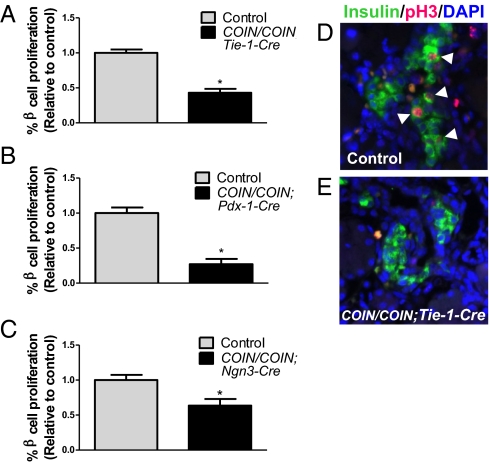
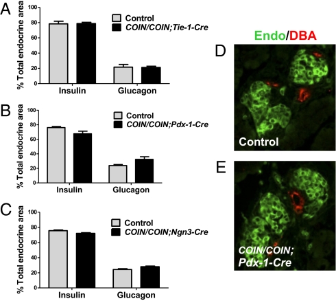
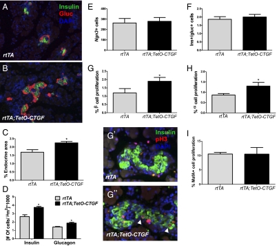
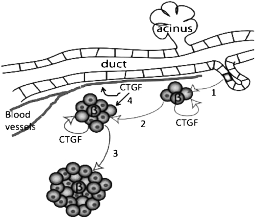
Type 1 and type 2 diabetes result from an absolute or relative reduction in functional β-cell mass. One approach to replacing lost β-cell mass is transplantation of cadaveric islets; however, this approach is limited by lack of adequate donor tissue. Therefore, there is much interest in identifying factors that enhance β-cell differentiation and proliferation in vivo or in vitro. Connective tissue growth factor (CTGF) is a secreted molecule expressed in endothelial cells, pancreatic ducts, and embryonic β cells that we previously showed is required for β-cell proliferation, differentiation, and islet morphogenesis during development. The current study investigated the tissue interactions by which CTGF promotes normal pancreatic islet development. We found that loss of CTGF from either endothelial cells or β cells results in decreased embryonic β-cell proliferation, making CTGF unique as an identified β cell-derived factor that regulates embryonic β-cell proliferation. Endothelial CTGF inactivation was associated with decreased islet vascularity, highlighting the proposed role of endothelial cells in β-cell proliferation. Furthermore, CTGF overexpression in β cells during embryogenesis using an inducible transgenic system increased islet mass at birth by promoting proliferation of immature β cells, in the absence of changes in islet vascularity. Together, these findings demonstrate that CTGF acts in an autocrine manner during pancreas development and suggest that CTGF has the potential to enhance expansion of immature β cells in directed differentiation or regeneration protocols.
Conflict of interest statement
Conflict of interest statement: A.N.E. is a paid employee and shareholder of Regeneron Pharmaceuticals.
Figures
Similar articles
-
Connective tissue growth factor (CTGF) inactivation leads to defects in islet cell lineage allocation and beta-cell proliferation during embryogenesis.Mol Endocrinol. 2009 Mar;23(3):324-36. doi: 10.1210/me.2008-0045. Epub 2009 Jan 8. Mol Endocrinol. 2009. PMID: 19131512 Free PMC article.
-
Connective tissue growth factor is critical for proper β-cell function and pregnancy-induced β-cell hyperplasia in adult mice.Am J Physiol Endocrinol Metab. 2016 Sep 1;311(3):E564-74. doi: 10.1152/ajpendo.00194.2016. Epub 2016 Jul 26. Am J Physiol Endocrinol Metab. 2016. Retraction in: Am J Physiol Endocrinol Metab. 2017 Jul 1;313(1):E105. doi: 10.1152/ajpendo.zh1-7764-retr.2017. PMID: 27460898 Free PMC article. Retracted.
-
Vascular-derived connective tissue growth factor (Ctgf) is critical for pregnancy-induced β cell hyperplasia in adult mice.Islets. 2017 Nov 2;9(6):150-158. doi: 10.1080/19382014.2017.1356963. Epub 2017 Nov 7. Islets. 2017. PMID: 29111856 Free PMC article.
-
Differential regulation of embryonic and adult β cell replication.Cell Cycle. 2012 Jul 1;11(13):2431-42. doi: 10.4161/cc.20545. Epub 2012 Jul 1. Cell Cycle. 2012. PMID: 22659844 Free PMC article. Review.
-
Regulation of pancreatic function by connective tissue growth factor (CTGF, CCN2).Cytokine Growth Factor Rev. 2013 Feb;24(1):59-68. doi: 10.1016/j.cytogfr.2012.07.001. Epub 2012 Aug 10. Cytokine Growth Factor Rev. 2013. PMID: 22884427 Free PMC article. Review.
Cited by
-
A physiological role for connective tissue growth factor in early wound healing.Lab Invest. 2013 Jan;93(1):81-95. doi: 10.1038/labinvest.2012.162. Epub 2012 Nov 19. Lab Invest. 2013. PMID: 23212098 Free PMC article.
-
Increased CCN2, substance P and tissue fibrosis are associated with sensorimotor declines in a rat model of repetitive overuse injury.J Cell Commun Signal. 2015 Mar;9(1):37-54. doi: 10.1007/s12079-015-0263-0. Epub 2015 Jan 24. J Cell Commun Signal. 2015. PMID: 25617052 Free PMC article.
-
Pancreas lineage allocation and specification are regulated by sphingosine-1-phosphate signalling.PLoS Biol. 2017 Mar 1;15(3):e2000949. doi: 10.1371/journal.pbio.2000949. eCollection 2017 Mar. PLoS Biol. 2017. PMID: 28248965 Free PMC article.
-
Macrophages are essential for CTGF-mediated adult β-cell proliferation after injury.Mol Metab. 2015 May 19;4(8):584-91. doi: 10.1016/j.molmet.2015.05.002. eCollection 2015 Aug. Mol Metab. 2015. PMID: 26266091 Free PMC article.
-
The Importance of Intra-Islet Communication in the Function and Plasticity of the Islets of Langerhans during Health and Diabetes.Int J Mol Sci. 2024 Apr 6;25(7):4070. doi: 10.3390/ijms25074070. Int J Mol Sci. 2024. PMID: 38612880 Free PMC article. Review.
References
-
- Apelqvist A, et al. Notch signalling controls pancreatic cell differentiation. Nature. 1999;400:877–881. - PubMed
-
- Jensen J, et al. Independent development of pancreatic alpha- and beta-cells from neurogenin3-expressing precursors: A role for the notch pathway in repression of premature differentiation. Diabetes. 2000;49(2):163–176. - PubMed
-
- Gu G, Dubauskaite J, Melton DA. Direct evidence for the pancreatic lineage: NGN3+ cells are islet progenitors and are distinct from duct progenitors. Development. 2002;129:2447–2457. - PubMed
-
- Bhushan A, et al. Fgf10 is essential for maintaining the proliferative capacity of epithelial progenitor cells during early pancreatic organogenesis. Development. 2001;128:5109–5117. - PubMed
Publication types
MeSH terms
Substances
Grants and funding
LinkOut - more resources
Full Text Sources
Molecular Biology Databases
Miscellaneous
