

First-ever Ischemic Stroke after a Flight in a Patient with Prior Poliomyelitis
- PMID: 21876645
- PMCID: PMC3160001
- DOI: 10.4137/cpath.s476
First-ever Ischemic Stroke after a Flight in a Patient with Prior Poliomyelitis
Abstract
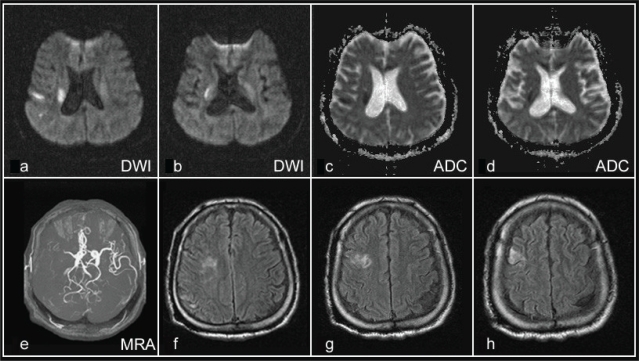
Survivors of poliomyelitis sometimes travel by air with mobility assistance. However, prolonged seating during long-haul flights may also possibly produce stroke events on polio-inflicted patients. A 48-year-old polio-inflicted male suffered a stroke after an extended flight. A two-dimensional echocardiography was normal without detected patent foramen ovale or dyskinetic segment. The venodynamic variables were all within normal limits. MR Imaging studies revealed acute cerebral infarction in the distribution of the right middle cerebral artery and posterior watershed area. Hematological examination revealed positive anti-cardiolipin IgG antibody which might contribute to the risk of thrombosis as an underlying condition in addition to immobilization. This is the first presentation of ischemic stroke after a flight in a patient with prior poliomyelitis. In addition to decompression sickness, economy class stroke syndrome and postpoliomyelitis syndrome, the physician should also take other coagulation disorders into consideration during the investigation.
Keywords: anticardiolipin antibodies; cabin pressurization; decompression sickness; dehydration; ischemic stroke; poliomyelitis.
Figures

Similar articles
-
Stroke and pulmonary thromboembolism after a long flight.Eur J Neurol. 2005 Sep;12(9):732-4. doi: 10.1111/j.1468-1331.2005.01070.x. Eur J Neurol. 2005. PMID: 16128878
-
Economy class stroke syndrome: case report and review of the literature.Eur J Vasc Endovasc Surg. 2004 Mar;27(3):239-43. doi: 10.1016/j.ejvs.2003.12.024. Eur J Vasc Endovasc Surg. 2004. PMID: 14760590 Review.
-
Comparison of diagnostic techniques for the detection of a patent foramen ovale in stroke patients.Stroke. 1993 Jul;24(7):1020-4. doi: 10.1161/01.str.24.7.1020. Stroke. 1993. PMID: 8322376
-
[Prevalence of patent foramen ovale in young patients with cerebral ischemic accident of unknown origin].Rev Esp Cardiol. 2003 Jul;56(7):662-8. doi: 10.1016/s0300-8932(03)76936-x. Rev Esp Cardiol. 2003. PMID: 12855148 Spanish.
-
The secret enemy during a flight: Economy class syndrome.Anatol J Cardiol. 2021 Aug;25(Suppl 1):13-17. doi: 10.5152/AnatolJCardiol.2021.S106. Anatol J Cardiol. 2021. PMID: 34464293 Free PMC article. Review.
References
-
- Aksoy FG. MR imaging of subclinical cerebral decompression sickness. A case report. Acta Radiol. 2003;44:108–10. - PubMed
-
- Carhuapoma JR, Mitsias P, Levine SR. Cerebral Venous Thrombosis and Anticardiolipin Antibodies. Stroke. 1997;28:2363–9. - PubMed
-
- Chen WH, Kao YF, Lan MY, et al. The Increase of Blood Anti-cardiolipin Antibody Depends on the Underlying Etiology in Cerebral Ischemia. Clin Appl Thromb Hemost. 2006;12:69–76. - PubMed
-
- D’Agati V, Kunis C, Williams G, et al. Anti-cardiolipin antibody and renal disease: a report of three cases. J Am Soc Nephrol. 1990;1:777–84. - PubMed
Publication types
LinkOut - more resources
Full Text Sources
