

The impact of bioactive lipids on cardiovascular development
- PMID: 21876704
- PMCID: PMC3159013
- DOI: 10.4061/2011/916180
The impact of bioactive lipids on cardiovascular development
Abstract
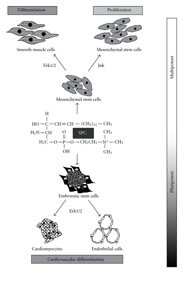
Lysophospholipids comprise a group of bioactive molecules with multiple biological functions. The cardinal members of this signalling molecule group are sphingosylphosphorylcholine (SPC), lysophosphatidic acid (LPA), and sphingosine 1-phosphate (S1P) which are, at least in part, homologous to each other. Bioactive lipids usually act via G-protein coupled receptors (GPCRs), but can also function as direct intracellular messengers. Recently, it became evident that bioactive lipids play a role during cellular differentiation development. SPC induces mesodermal differentiation of mouse ES cells and differentiation of promyelocytic leukemia cells, by a mechanism being critically dependent on MEK-ERK signalling. LPA stimulates the clonal expansion of neurospheres from neural stem/progenitor cells and induces c-fos via activation of mitogen- and stress-activated protein kinase 1 (MSK1) in ES cells. S1P acts on hematopoietic progenitor cells as a chemotactic factor and has also been found to be critical for cardiac and skeletal muscle regeneration. Furthermore, S1P promotes cardiogenesis and similarly activates Erk signalling in mouse ES cells. Interestingly, S1P may also act to maintain human stem cell pluripotency. Both LPA and S1P positively regulate the proliferative capacity of murine ES cells. In this paper we will focus on the differential and developmental impact of lysophospholipids on cardiovascular development.
Figures


Similar articles
-
Convergent regulation of neuronal differentiation and Erk and Akt kinases in human neural progenitor cells by lysophosphatidic acid, sphingosine 1-phosphate, and LIF: specific roles for the LPA1 receptor.ASN Neuro. 2014 Nov 24;6(6):1759091414558416. doi: 10.1177/1759091414558416. Print 2014. ASN Neuro. 2014. PMID: 25424429 Free PMC article.
-
The bioactive lipid sphingosylphosphorylcholine induces differentiation of mouse embryonic stem cells and human promyelocytic leukaemia cells.Cell Signal. 2007 Feb;19(2):367-77. doi: 10.1016/j.cellsig.2006.07.015. Epub 2006 Jul 28. Cell Signal. 2007. PMID: 16978842
-
Lysophospholipid receptors: signalling, pharmacology and regulation by lysophospholipid metabolism.Biochim Biophys Acta. 2007 Apr;1768(4):923-40. doi: 10.1016/j.bbamem.2006.09.026. Epub 2006 Oct 4. Biochim Biophys Acta. 2007. PMID: 17078925 Review.
-
Bioactive lipids lysophosphatidic acid and sphingosine 1-phosphate mediate breast cancer cell biological functions through distinct mechanisms.Oncol Res. 2009;18(4):173-84. doi: 10.3727/096504009790217399. Oncol Res. 2009. PMID: 20112503 Free PMC article.
-
Novel implications for lysophospholipids, lysophosphatidic acid and sphingosine 1-phosphate, as drug targets in cancer.Anticancer Agents Med Chem. 2009 May;9(4):381-91. doi: 10.2174/1871520610909040381. Anticancer Agents Med Chem. 2009. PMID: 19442039 Review.
Cited by
-
Critical Role of the Sphingolipid Pathway in Stroke: a Review of Current Utility and Potential Therapeutic Targets.Transl Stroke Res. 2016 Oct;7(5):420-38. doi: 10.1007/s12975-016-0477-3. Epub 2016 Jun 24. Transl Stroke Res. 2016. PMID: 27339463 Free PMC article. Review.
-
Stage-specific Effects of Bioactive Lipids on Human iPSC Cardiac Differentiation and Cardiomyocyte Proliferation.Sci Rep. 2018 Apr 26;8(1):6618. doi: 10.1038/s41598-018-24954-3. Sci Rep. 2018. PMID: 29700394 Free PMC article.
-
Bioactive Lipid Signaling in Cardiovascular Disease, Development, and Regeneration.Cells. 2020 Jun 3;9(6):1391. doi: 10.3390/cells9061391. Cells. 2020. PMID: 32503253 Free PMC article. Review.
-
Revisiting the role of lysophosphatidic acid in stem cell biology.Exp Biol Med (Maywood). 2021 Aug;246(16):1802-1809. doi: 10.1177/15353702211019283. Epub 2021 May 26. Exp Biol Med (Maywood). 2021. PMID: 34038224 Free PMC article. Review.
-
Definitive Endoderm Formation from Plucked Human Hair-Derived Induced Pluripotent Stem Cells and SK Channel Regulation.Stem Cells Int. 2013;2013:360573. doi: 10.1155/2013/360573. Epub 2013 Apr 16. Stem Cells Int. 2013. PMID: 23710194 Free PMC article.
References
-
- Rodriguez-Lafrasse C, Vanier MT. Sphingosylphosphorylcholine in Niemann-Pick disease brain: accumulation in type A but not in type B. Neurochemical Research. 1999;24(2):199–205. - PubMed
-
- Meyer zu Heringdorf D, Himmel HM, Jakobs KH. Sphingosylphosphorylcholine—biological functions and mechanisms of action. Biochimica et Biophysica Acta. 2002;1582(1–3):178–189. - PubMed
-
- Murata N, Sato K, Kon J, Tomura H, Okajima F. Quantitative measurement of sphingosine 1-phosphate by radioreceptor- binding assay. Analytical Biochemistry. 2000;282(1):115–120. - PubMed
-
- Hara J, Higuchi K, Okamoto R, Kawashima M, Imokawa G. High-expression of sphingomyelin deacylase is an important determinant of ceramide deficiency leading to barrier disruption in atopic dermatitis. Journal of Investigative Dermatology. 2000;115(3):406–413. - PubMed
LinkOut - more resources
Full Text Sources
Other Literature Sources
Research Materials
Miscellaneous
