

Efficient and scalable purification of cardiomyocytes from human embryonic and induced pluripotent stem cells by VCAM1 surface expression
- PMID: 21876760
- PMCID: PMC3158088
- DOI: 10.1371/journal.pone.0023657
Efficient and scalable purification of cardiomyocytes from human embryonic and induced pluripotent stem cells by VCAM1 surface expression
Abstract
Rationale: Human embryonic and induced pluripotent stem cells (hESCs/hiPSCs) are promising cell sources for cardiac regenerative medicine. To realize hESC/hiPSC-based cardiac cell therapy, efficient induction, purification, and transplantation methods for cardiomyocytes are required. Though marker gene transduction or fluorescent-based purification methods have been reported, fast, efficient and scalable purification methods with no genetic modification are essential for clinical purpose but have not yet been established. In this study, we attempted to identify cell surface markers for cardiomyocytes derived from hESC/hiPSCs.
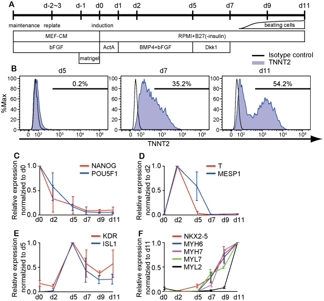
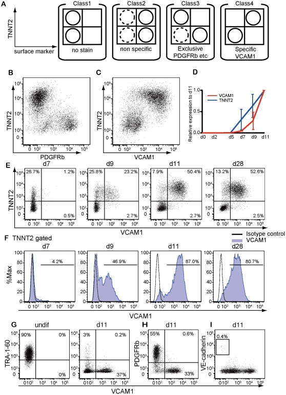
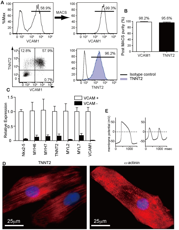
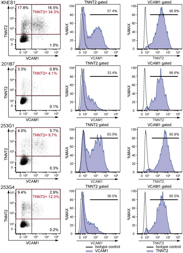
Method and result: We adopted a previously reported differentiation protocol for hESCs based on high density monolayer culture to hiPSCs with some modification. Cardiac troponin-T (TNNT2)-positive cardiomyocytes appeared robustly with 30-70% efficiency. Using this differentiation method, we screened 242 antibodies for human cell surface molecules to isolate cardiomyocytes derived from hiPSCs and identified anti-VCAM1 (Vascular cell adhesion molecule 1) antibody specifically marked cardiomyocytes. TNNT2-positive cells were detected at day 7-8 after induction and 80% of them became VCAM1-positive by day 11. Approximately 95-98% of VCAM1-positive cells at day 11 were positive for TNNT2. VCAM1 was exclusive with CD144 (endothelium), CD140b (pericytes) and TRA-1-60 (undifferentiated hESCs/hiPSCs). 95% of MACS-purified cells were positive for TNNT2. MACS purification yielded 5-10×10(5) VCAM1-positive cells from a single well of a six-well culture plate. Purified VCAM1-positive cells displayed molecular and functional features of cardiomyocytes. VCAM1 also specifically marked cardiomyocytes derived from other hESC or hiPSC lines.
Conclusion: We succeeded in efficiently inducing cardiomyocytes from hESCs/hiPSCs and identifying VCAM1 as a potent cell surface marker for robust, efficient and scalable purification of cardiomyocytes from hESC/hiPSCs. These findings would offer a valuable technological basis for hESC/hiPSC-based cell therapy.
Conflict of interest statement
Figures
References
-
- Laflamme MA, Chen KY, Naumova AV, Muskheli V, Fugate JA, et al. Cardiomyocytes derived from human embryonic stem cells in pro-survival factors enhance function of infarcted rat hearts. Nat Biotechnol. 2007;25:1015–1024. - PubMed
-
- Yang L, Soonpaa MH, Adler ED, Roepke TK, Kattman SJ, et al. Human cardiovascular progenitor cells develop from a KDR+ embryonic-stem-cell-derived population. Nature. 2008;453:524–528. - PubMed
-
- Kattman SJ, Witty AD, Gagliardi M, Dubois NC, Niapour M, et al. Stage-Specific Optimization of Activin/Nodal and BMP Signaling Promotes Cardiac Differentiation of Mouse and Human Pluripotent Stem Cell Lines. Cell Stem Cell. 2011;8:228–240. - PubMed
-
- Narazaki G, Uosaki H, Teranishi M, Okita K, Kim B, et al. Directed and systematic differentiation of cardiovascular cells from mouse induced pluripotent stem cells. Circulation. 2008;118:498–506. - PubMed
Publication types
MeSH terms
Substances
LinkOut - more resources
Full Text Sources
Other Literature Sources
Research Materials
Miscellaneous
