. 2011;6(8):e23673.
doi: 10.1371/journal.pone.0023673. Epub 2011 Aug 18.

A signature in HIV-1 envelope leader peptide associated with transition from acute to chronic infection impacts envelope processing and infectivity

Affiliations

A signature in HIV-1 envelope leader peptide associated with transition from acute to chronic infection impacts envelope processing and infectivity

Mohammed Asmal et al. PLoS One. 2011.

Abstract

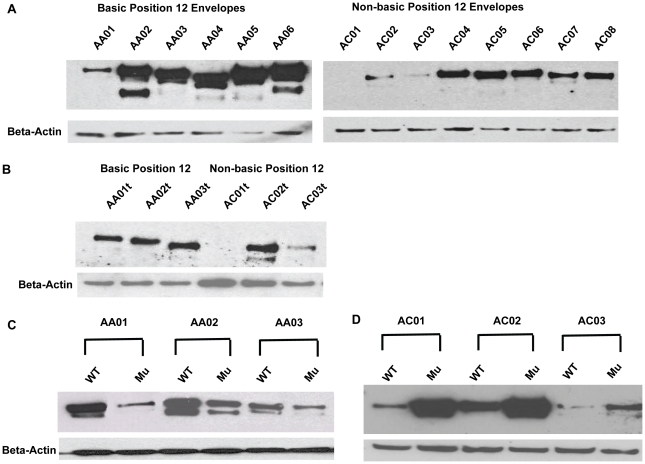
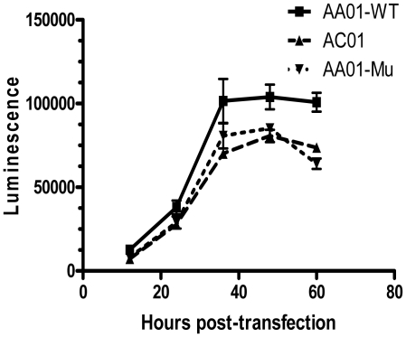
Mucosal transmission of the human immunodeficiency virus (HIV) results in a bottleneck in viral genetic diversity. Gnanakaran and colleagues used a computational strategy to identify signature amino acids at particular positions in Envelope that were associated either with transmitted sequences sampled very early in infection, or sequences sampled during chronic infection. Among the strongest signatures observed was an enrichment for the stable presence of histidine at position 12 at transmission and in early infection, and a recurrent loss of histidine at position 12 in chronic infection. This amino acid lies within the leader peptide of Envelope, a region of the protein that has been shown to influence envelope glycoprotein expression and virion infectivity. We show a strong association between a positively charged amino acid like histidine at position 12 in transmitted/founder viruses with more efficient trafficking of the nascent envelope polypeptide to the endoplasmic reticulum and higher steady-state glycoprotein expression compared to viruses that have a non-basic position 12 residue, a substitution that was enriched among viruses sampled from chronically infected individuals. When expressed in the context of other viral proteins, transmitted envelopes with a basic amino acid position 12 were incorporated at higher density into the virus and exhibited higher infectious titers than did non-signature envelopes. These results support the potential utility of using a computational approach to examine large viral sequence data sets for functional signatures and indicate the importance of Envelope expression levels for efficient HIV transmission.


Conflict of interest statement

Competing Interests: The authors have declared that no competing interests exist.

Figures

Figure 1
Figure 1. Maximum likelihood phylogenetic tree for envelope sequences used in this study.
Sequence alignments and ML tree were generated using Seaview 4.0 . Envelopes with the basic position 12 residue are boxed and those lacking the signature are not boxed.
Figure 2
Figure 2. Position 12 signature enhances steady-state envelope expression in Jurkat cells.
(A) Transmitted envelopes were expressed in Jurkat cells by transient transfection and cell lysates analyzed by SDS-PAGE and Western blot, probed with the 3B3 monoclonal anti-gp120 antibody. Equal well loading was confirmed by subsequent membrane probing with a monoclonal antibody to beta-actin. Envelope signal intensities were quantified by Quantity One (BioRad). Mean signal for AA01–AA06: 3433 arbitrary units, standard deviation: 973. Mean and standard deviation for AC01–AC08: 1379 and 481, respectively. P value by Students' two-tailed T-test = .0002 for difference between the two groups. (B) Six transmitted envelopes were carboxy-terminus V5 epitope tagged and expressed in Jurkat cells. Western blots were probed with a monoclonal antibody to V5. (C) Single point mutations were introduced into the sequences of three envelopes, converting the native histidine or arginine (WT) into non-basic glutamine (Mu). Mean decrement in protein expression caused by position 12 mutation was 61%, with a standard deviation of 16% among the three envelopes analyzed. (D) Single point mutations were introduced into three envelopes converting the native glutamine or alignment gap (WT) into basic histidine (Mu). Mean increase in protein expression caused by position 12 mutation was 3.6 fold with a standard deviation of 1.9 fold.
Figure 3
Figure 3. Position 12 polymorphism influences efficiency of leader peptide in regulating protein transport through the secretory pathway.
Secreted luciferase reporter constructs were generated to help quantify leader peptide efficiency. Fusion PCR was utilized to substitute the 31 base pair leader peptide of HIV-1 envelope for the 18 base pair Metridia longa secreted luciferase leader peptide. Secreted luciferase constructs were generated bearing the leader peptides of envelopes AA01, AC01 and AA01-Mu containing the histidine to glutamine mutation. These were co-transfected in triplicate into Jurkat cells with a human secreted alkaline phosphatase control vector (pSEAP2, Clontech). Culture supernatants were collected at 12, 24, 48, 36 and 60 hours post-transfection, and analyzed in parallel for luciferase and SEAP activity. Luciferase activity was normalized to the SEAP control, and mean and standard deviation of triplicate transfection results are plotted. Two-tailed Student's T test for comparison of AA01 to AC01 (p = 0.0171) and AA01 to AA01-mu (p = 0.0318) were significant, while comparison of AC01 to AA01-mu revealed no significant difference.
Figure 4
Figure 4. Pseudovirions bearing leader peptide signature envelopes are more infectious in a single round infectivity assay.
HIV-1 pseudovirions were generated by co-transfection of 293T cells with pcDNA 3.1 transmitted envelopes and SG3deltaEnv, a plasmid encoding all HIV-1 structural and accessory proteins except for envelope. SG3deltaEnv and the envelope constructs were transfected at a molar ratio of approximately four to one. Culture supernatants were collected 24 to 48 hours after transfection, and passed through a 0.2 micron filter. Five-fold serial dilutions of supernatants were incubated in 96-well plates with 2×10⁴ TZM-bl cells, a reporter cell line expressing HIV-1 co-receptors as well as a Tat-sensitive luciferase reporter. Pseudovirus and TZM-bl cells were incubated for 48 hours before luciferase activity was quantified. This experiment is representative of multiple studies. Six basic (dashed lines) and eight non-basic (solid) transmitted envelopes were assayed; position 12 residue is indicated parenthetically. Relative luciferase activity at each dilution was compared between all basic and non-signature envelopes by Mann-Whitney test. Dilutions at which p-values for comparison are <.05 indicated by *, and <.01 indicated by **.
Figure 5
Figure 5. Position 12 signature is associated with increased envelope incorporation into pseudovirions.
(A) Pseudovirions were generated in 293T cells, and culture supernatants were layered over 20% sucrose and ultracentrifuged at 27,000 RPM for 2 hours. P24 was quantified by ELISA (Zeptometrix). (B) Pelleted pseudovirions were normalized for p24, and subsequently analyzed by Western blot using the 3B3 antibody.
Figure 6
Figure 6. Precise quantification of p24 and envelope content in pseudovirions.
Fifty milliliter volumes of 293T pseudovirion supernatants were generated for Envelopes AA01 and AC01. Supernatants were pelleted over a sucrose cushion, and resuspended in PBS. Equal volumes of resuspended AA01 and AC01 were analyzed by SDS-PAGE. Varying dilutions of a previously quantified virus were run simultaneously. Protein signal was measured by densitometry, and p24 and envelope in AA01 and AC01 pseudovirus were quantified by comparison to dilutions of known virus. The ratio of p24 to envelope was calculated for both samples.
Figure 7
Figure 7. Alteration of ratio of pseudovirion p24:envelope does not alter differential pseudovirion incorporation of envelope between signature and non-signature envelopes.
Position 12 histidine bearing envelope AA05 and non-histidine bearing envelope AC02 were co-transfected with SG3deltaEnv at two different ratios of plasmid DNA to generate pseudovirions in 293T cells. Pseudovirions were purified by ultracentrifugation over 20% sucrose. Pellets were resupended and analyzed by Western Blot for envelope content and by p24 ELISA for Gag content. Pelleted pseudovirus was also applied to TZM-bl reporter cells and luciferase activity was measured as an indication of infectivity.
Figure 8
Figure 8. Mathematical modeling of the role of viral infectivity in acute and chronic infection.
(A) The exponential growth rate of virus during early infection was plotted as a function of viral infectivity, k, using the equation: r0 = (k p/c)T0 – δ. Estimates for p, c, T0 and δ were taken from Stafford, et al. . (B) Viral load at steady-state was plotted as a function of viral infectivity, k, using the equation: Vss = (πλ)/(δc) – d/k, also taken from Stafford, et al. and Nowak, et al. .

References

    1. Learn GH, Muthui D, Brodie SJ, Zhu T, Diem K, et al. Virus population homogenization following acute human immunodeficiency virus type 1 infection. J Virol. 2002;76:11953–11959. - PMC - PubMed
    1. McNearney T, Hornickova Z, Markham R, Birdwell A, Arens M, et al. Relationship of human immunodeficiency virus type 1 sequence heterogeneity to stage of disease. Proc Natl Acad Sci U S A. 1992;89:10247–10251. - PMC - PubMed
    1. Wolfs TF, Zwart G, Bakker M, Goudsmit J. HIV-1 genomic RNA diversification following sexual and parenteral virus transmission. Virology. 1992;189:103–110. - PubMed
    1. Zhu T, Mo H, Wang N, Nam DS, Cao Y, et al. Genotypic and phenotypic characterization of HIV-1 patients with primary infection. Science. 1993;261:1179–1181. - PubMed
    1. Frater AJ, Edwards CT, McCarthy N, Fox J, Brown H, et al. Passive sexual transmission of human immunodeficiency virus type 1 variants and adaptation in new hosts. J Virol. 2006;80:7226–7234. - PMC - PubMed

Publication types

MeSH terms