. 2011:2011:741723.
doi: 10.4061/2011/741723. Epub 2011 Aug 22.

Molecular beacons: powerful tools for imaging RNA in living cells

Affiliations

Molecular beacons: powerful tools for imaging RNA in living cells

Ricardo Monroy-Contreras et al. J Nucleic Acids. 2011.

Abstract

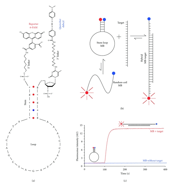
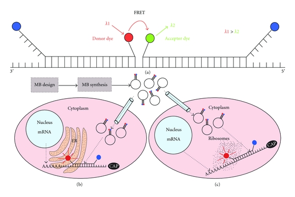
Recent advances in RNA functional studies highlights the pivotal role of these molecules in cell physiology. Diverse methods have been implemented to measure the expression levels of various RNA species, using either purified RNA or fixed cells. Despite the fact that fixed cells offer the possibility to observe the spatial distribution of RNA, assays with capability to real-time monitoring RNA transport into living cells are needed to further understand the role of RNA dynamics in cellular functions. Molecular beacons (MBs) are stem-loop hairpin-structured oligonucleotides equipped with a fluorescence quencher at one end and a fluorescent dye (also called reporter or fluorophore) at the opposite end. This structure permits that MB in the absence of their target complementary sequence do not fluoresce. Upon binding to targets, MBs emit fluorescence, due to the spatial separation of the quencher and the reporter. Molecular beacons are promising probes for the development of RNA imaging techniques; nevertheless much work remains to be done in order to obtain a robust technology for imaging various RNA molecules together in real time and in living cells. The present work concentrates on the different requirements needed to use successfully MB for cellular studies, summarizing recent advances in this area.


Figures

Figure 1
Figure 1
Structure and function of MB. (a) Stem-loop hairpin structure of a MB showing its four structural components: loop, stem, quencher, and reporter. The chemical structure of the linkers is drawn according to the manufacturer (Integrated DNA Technologies, Iowa, USA). (b) Mechanism of MB function. A MB in a solution containing both MB and target could be in three states: free in a stem-loop hairpin conformation, hybridized with it target, or unbound in a random-coil conformation. The random-coil conformation of the MB contributes to the background. (c) Fluorescence intensity. The blue line shows the fluorescence intensity for a MB in solution (50 nM) during 400 seconds (background), and the red line corresponds to the addition (100 seconds) of a target oligonucleotide (500 nM) that hybridize the MB at the loop region. An increase in fluorescent intensity of approximately thirteenfold is observed. The reporter is represented with a red circle and the quencher with a blue. UA means arbitrary units.
Figure 2
Figure 2
Different positions for MB-target hybridization. The loop region is illustrated in green and the stem in purple for all panels, target is brown and the bonds MB target are black ((b), (c), (d) y (e)). (a) MB with a stem of five nucleotides in the stem-loop hairpin conformation. (b) MB with a stem of five nucleotides and twenty two nucleotides in the loop, target hybridization occurs only at loop region. (c) MB with a stem of five nucleotides and a stem of seventeen nucleotides, using the loop and all the 3′ arm to hybridize the target (3′ shared stem MB). (d) MB with a stem of five nucleotides and a loop of seventeen nucleotides, using the loop and completely the 5′ arm to hybridize its target (5′ shared stem MB). (e) MB with a stem of five nucleotides and loop of eighteen nucleotides using partially both arms to hybridize its target (two nucleotides of every arm). Notice that a target that makes possible the design of a MB that hybridizes it using completely both arms could have a strong secondary structure that makes impossible the MB-target hybridization.
Figure 3
Figure 3
Structures of the superquenched MB. MB with one (1Q), two (2Q), and three (3Q) Dabcyl quenchers. The structures are represented in their oxidized state. The linker structures, the reporter linked to the 3′ end and the quencher or quenchers attached at the 5′ terminus correspond to [19].
Figure 4
Figure 4
MB linked to penetrating peptides. (a) Reaction of conjugation among the maleimide molecule with a carrier group (R1) and the thiol group of another molecule. This reaction may possibly link MB-peptide and occurs at a pH between 6.5–7.5. (b) One possibility using the maleimide-thiol system is that the peptide (NLS or CCP) it is linked to the maleimide across the secondary amine and reacts with a sulfhydryl group at the terminus of the MB hydrocarbon linker. (b′) The other possibility for the maleimide-thiol system is that the maleimide is linked at the terminus of the MB hydrocarbon linker and reacts with a thiol group at the peptide (NLS or CCP). (c) The link between the hydrocarbon linker of the MB and the peptide also could be across a disulphide bridge, in this case exists the possibility to cleave the bond in a reducing environment. (d) Using SLO to make permeable the cytoplasmic membrane is possible to introduce a MB linked to an NLS peptide to the cytoplasm with the objective that it be transported into the nucleus by the cellular machinery. For a CCP-linked MB the entrance to the nucleus is impossible.
Figure 5
Figure 5
Approaches for RNA visualization in living cells. (a) In order to reduce the possibility of false-positive signals and also increase the specificity, one can design two shared stem MB having a pair reporters compatible with FRET, one to serve like a donor dye (red circle) and the other like a acceptor dye (green circle). With this strategy the sequence to be recognized increases its length and thus the specificity of the assay (observe that one MB needs to have the reporter at the 3′ end and the quencher at the 5′ end, in a not conventional attachment). After the MB design and synthesis the probe could be delivered using microinjection; in conjunction with other MB or fluorescent dyes could be used to measure RNA distribution at subcellular level. For example it is possible to use a specific fluorescent dye for the endoplasmic reticulum (ER) and also a MB designed to hybridize a given sequence of the ribosome. If a particular mRNA (based on the fluorescence of the MB) colocalizes with the ER dye and ribosome dye it means that this mRNA is translated in ribosomes at ER (b) or if the mRNA signal colocalizes only with the ribosome dye means that the mRNA translation occurs at free ribosomes (c); also it is possible that the mRNA translation takes place at both free and ER associated ribosomes. These are only some examples of the power of using MB in combination with other compartment or structure-specific dyes.

References

    1. Glišin V, Crkvenjakov R, Byus C. Ribonucleic acid isolated by cesium chloride centrifugation. Biochemistry. 1974;13(12):2633–2637. - PubMed
    1. Auffray C, Rougeon F. Purification of mouse immunoglobulin heavy-chain messenger RNAs from total myeloma tumor RNA. European Journal of Biochemistry. 1980;107(2):303–314. - PubMed
    1. Chomczynski P, Sacchi N. Single-step method of RNA isolation by acid guanidinium thiocyanate-phenol-chloroform extraction. Analytical Biochemistry. 1987;162(1):156–159. - PubMed
    1. Saiki RK, Scharf S, Faloona F. Enzymatic amplification of β-globin genomic sequences and restriction site analysis for diagnosis of sickle cell anemia. Science. 1985;230(4732):1350–1354. - PubMed
    1. Alwine JC, Kemp DJ, Parker BA, et al. Detection of specific RNAs or specific fragments of DNA by fractionation in gels and transfer to diazobenzyloxymethyl paper. Methods in Enzymology. 1979;68:220–242. - PubMed

LinkOut - more resources