

Whole slide quantification of stromal lymphatic vessel distribution and peritumoral lymphatic vessel density in early invasive cervical cancer: a method description
- PMID: 21876817
- PMCID: PMC3163137
- DOI: 10.5402/2011/354861
Whole slide quantification of stromal lymphatic vessel distribution and peritumoral lymphatic vessel density in early invasive cervical cancer: a method description
Abstract
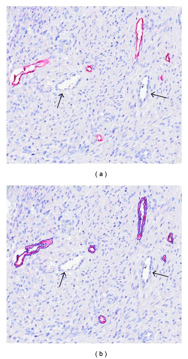
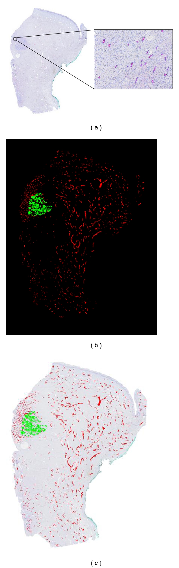
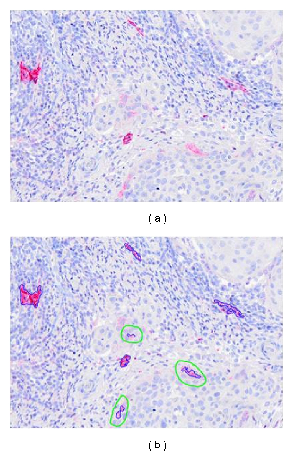
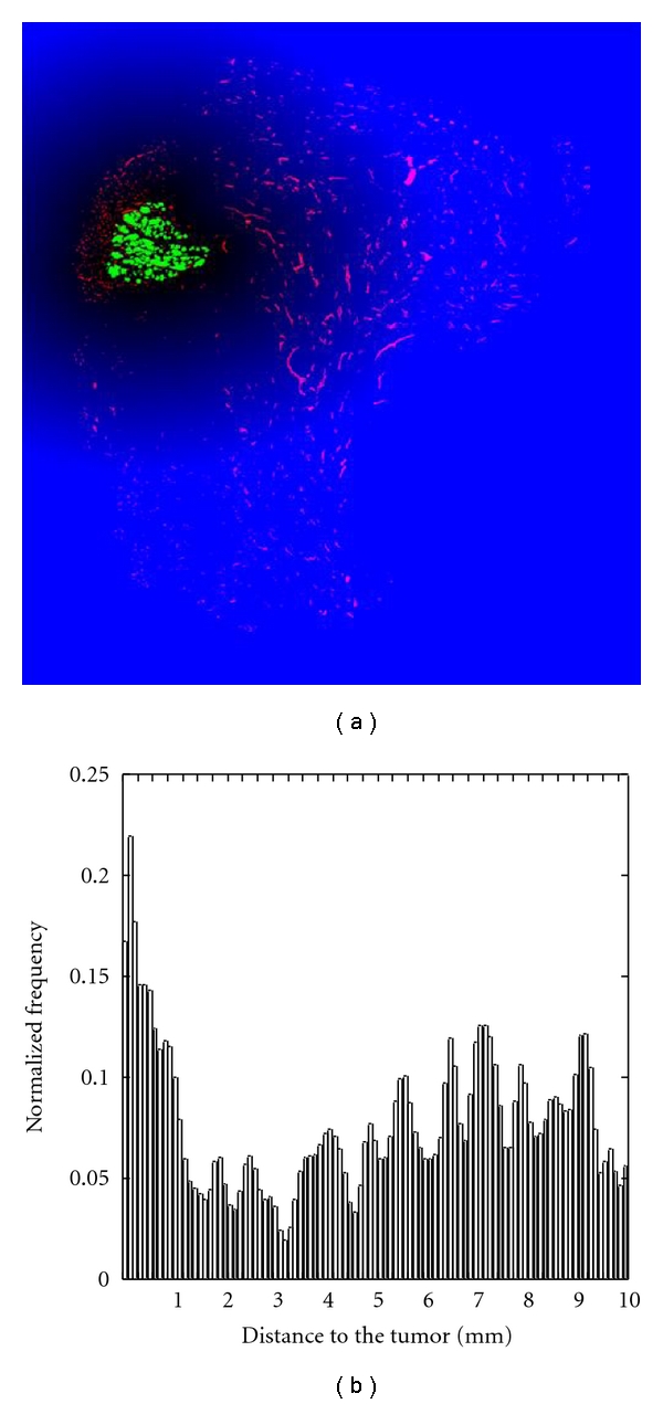
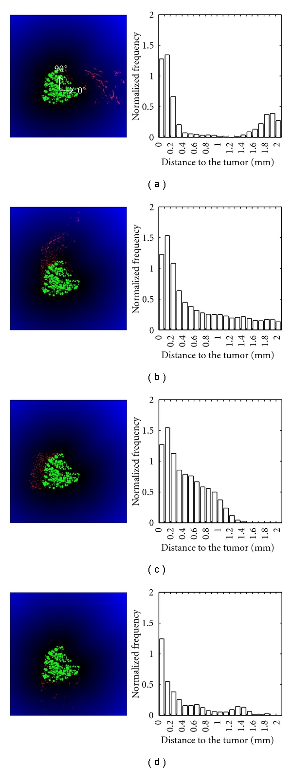
Peritumoral Lymphatic Vessel Density (LVD) is considered to be a predictive marker for the presence of lymph node metastases in cervical cancer. However, when LVD quantification relies on conventional optical microscopy and the hot spot technique, interobserver variability is significant and yields inconsistent conclusions. In this work, we describe an original method that applies computed image analysis to whole slide scanned tissue sections following immunohistochemical lymphatic vessel staining. This procedure allows to determine an objective LVD quantification as well as the lymphatic vessel distribution and its heterogeneity within the stroma surrounding the invasive tumor bundles. The proposed technique can be useful to better characterize lymphatic vessel interactions with tumor cells and could potentially impact on prognosis and therapeutic decisions.
Figures
References
-
- Delgado G, Bundy B, Zaino R, Sevin BU, Creasman WT, Major F. Prospective surgical-pathological study of disease-free interval in patients with stage IB squamous cell carcinoma of the cervix: a gynecologic oncology group study. Gynecologic Oncology. 1990;38(3):352–357. - PubMed
-
- Hacker NF, Wain GV, Nicklin JL. Resection of bulky positive lymph nodes in patients with cervical carcinoma. International Journal of Gynecological Cancer. 1995;5(4):250–256. - PubMed
-
- Trappen POV, Pepper MS. Lymphangiogenesis in human gynaecological cancers. Angiogenesis. 2005;8(2):137–145. - PubMed
-
- Sleeman JP, Thiele W. Tumor metastasis and the lymphatic vasculature. International Journal of Cancer. 2009;125(12):2747–2756. - PubMed
-
- Gombos Z, Xu X, Chu CS, Zhang PJ, Acs G. Peritumoral lymphatic vessel density and vascular endothelial growth factor C expression in early-stage squamous cell carcinoma of the uterine cervix. Clinical Cancer Research. 2005;11(23):8364–8371. - PubMed
LinkOut - more resources
Full Text Sources
