

doi: 10.4061/2011/739157.
Epub 2011 Aug 16.
CMR in Heart Failure
Affiliations
- PMID: 21876825
- PMCID: PMC3157673
- DOI: 10.4061/2011/739157
Item in Clipboard
CMR in Heart Failure
Cardiol Res Pract.
2011.
Abstract
Heart Failure (HF) is a common syndrome with multiple causes. Cardiovascular magnetic resonance (CMR) is a medical imaging technique with significant advantages, allowing the understanding of aetiology and pathophysiology of HF in the individual patient, permitting specific therapy to be administered and predicting prognosis. This paper discusses the diverse role of CMR in HF.
Figures
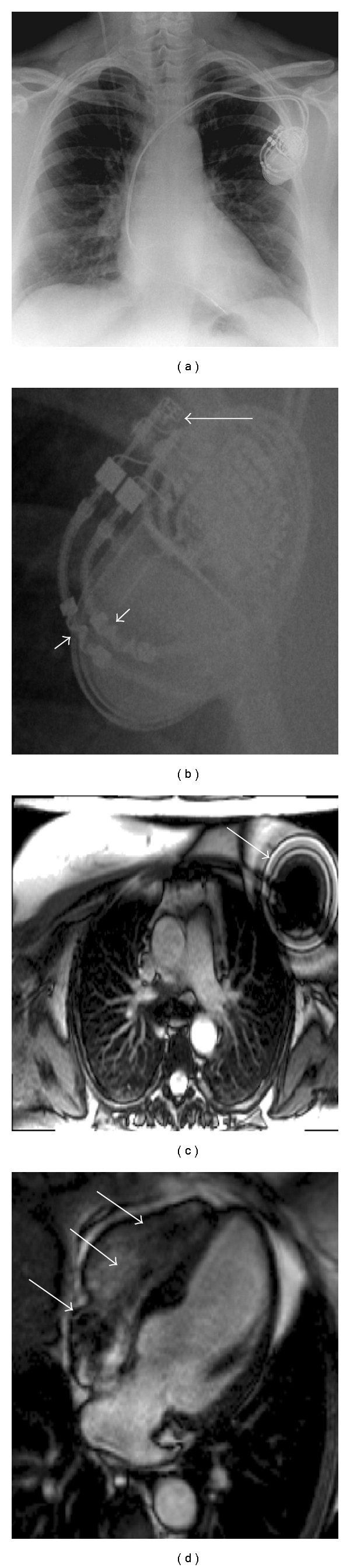
A CMR conditional pacemaker. (a, b) show the pacemaker on a chest X-ray. On (b) the arrows point to MRI conditional markers on the header and leads. (c) shows a large artifact from the pacemaker box on a transverse white blood stack. (d) shows suseptibility artifact from the pacemaker leads in the heart.
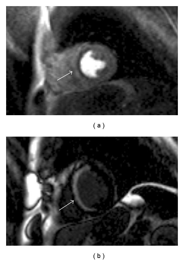
CMR in a patient with ischaemic cardiomyopathy. The cine imaging had shown normal wall thickness throughout but septal akinesis. (a) First pass perfusion sequence following administration of vasodilator stress with adenosine. A defect is seen throughout the septum. (b) Almost full thickness LGE is seen in the septum highly suggestive that this area is nonviable.
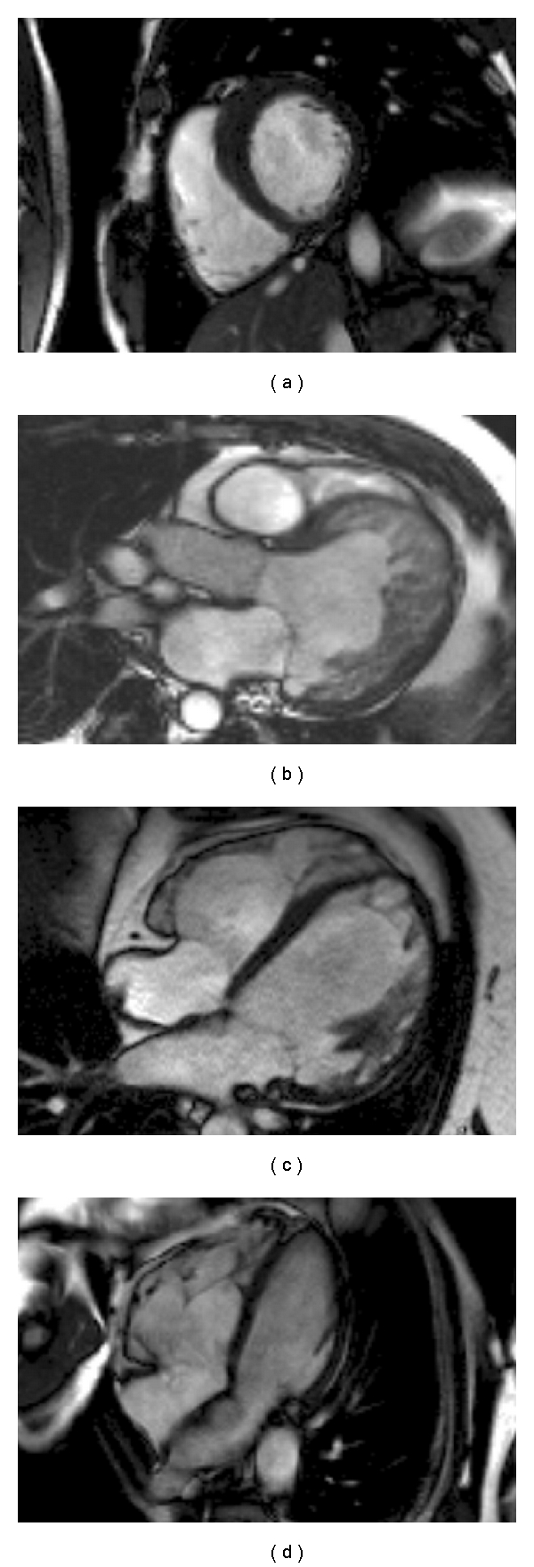
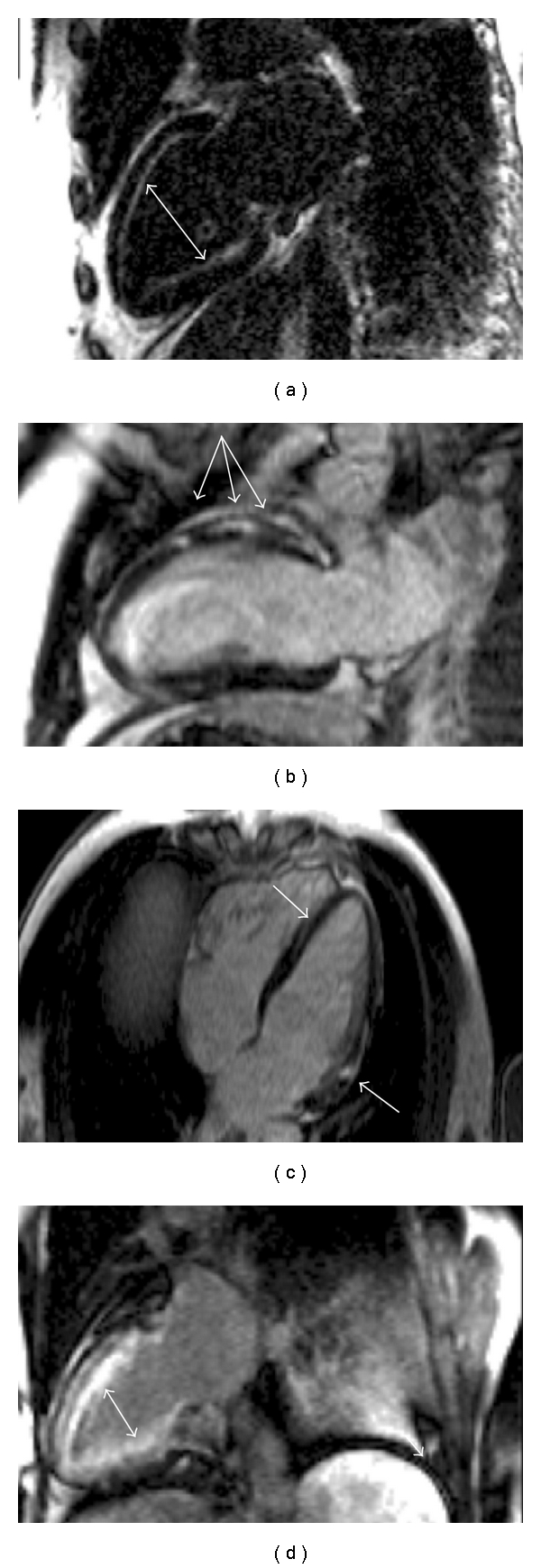
CMR cine sequences demonstrating the differing morphologies of inherited cardiomyopathies. (a) HCM: asymmetric basal anterospetal wall hypertrophy; (b) LVNC: left ventricular hypertrabeculation and dilatation; (c) FDCM: left ventricular wall thinning and dilatation; (d) ARVC: right ventricular dilatation.
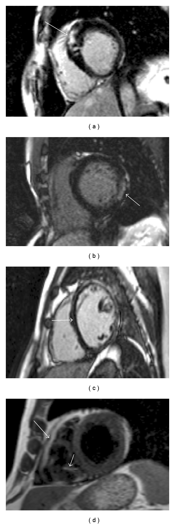
Tissue characterisation in inherited cardiomyopathy. (a) Patchy anteroseptal wall LGE in HCM; (b) Basal inferolateral wall LGE in Anderson Fabry Disease; (c) Septal and anterior mid wall LGE in familial FDCM; (d) RV fatty replacement in ARVC on the trabeculae and possibly the free wall.
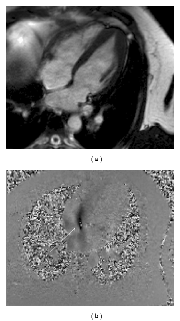
A patient with known apical HCM in whom transthoracic echocardiography had suggested a secundum ASD. (a) Cine 4 chamber image showing apical hypertrophy (b) flow sequence planned using the 4 chamber view shows flow across the intra atrial septum from left to right (arrowed), confirming the diagnosis. In this case, although the right heart does not appear dilated, the Qp : Qs was calculated as 2 showing the presence of a significant shunt.
Gadolinium-enhanced imaging in specific cardiac diseases. (a) Subendocardial LGE in a patient with AL amyloidosis. (b) Mid anterior wall focal LGE lesions in a patient with sarcoidosis. (c) Epicardial and mid wall anterior LGE in a patient with myocarditis. (d) Subendocardial enhancement in a patient with Churgg Strauss syndrome.
References
-
- Lloyd-Jones D, Adams RJ, Brown TM, et al. Executive summary: heart disease and stroke statistics-2010 update: a report from the american heart association. Circulation. 2010;121(7):e46–e215. - PubMed
-
- Bruder O, Schneider S, Nothnagel D, et al. EuroCMR (European Cardiovascular Magnetic Resonance) registry. Results of the german pilot phase. Journal of the American College of Cardiology. 2009;54(15):1457–1466. - PubMed
LinkOut - more resources
Full Text Sources
Research Materials
Miscellaneous