. 2011:2011:374286.
doi: 10.4061/2011/374286. Epub 2011 Aug 25.

Role of apoptosis in rabies viral encephalitis: a comparative study in mice, canine, and human brain with a review of literature

Affiliations

Role of apoptosis in rabies viral encephalitis: a comparative study in mice, canine, and human brain with a review of literature

M S Suja et al. Patholog Res Int. 2011.

Abstract

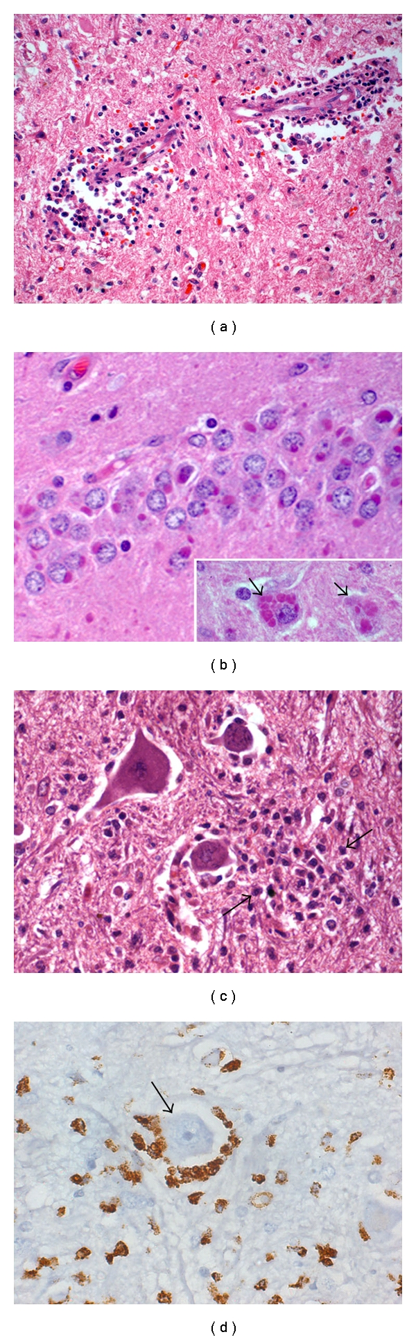
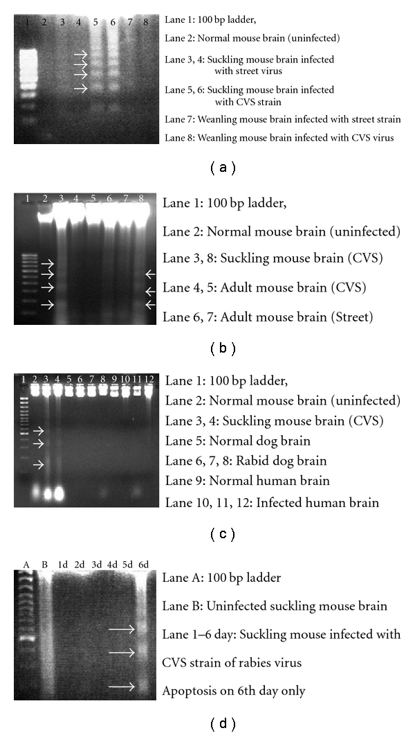
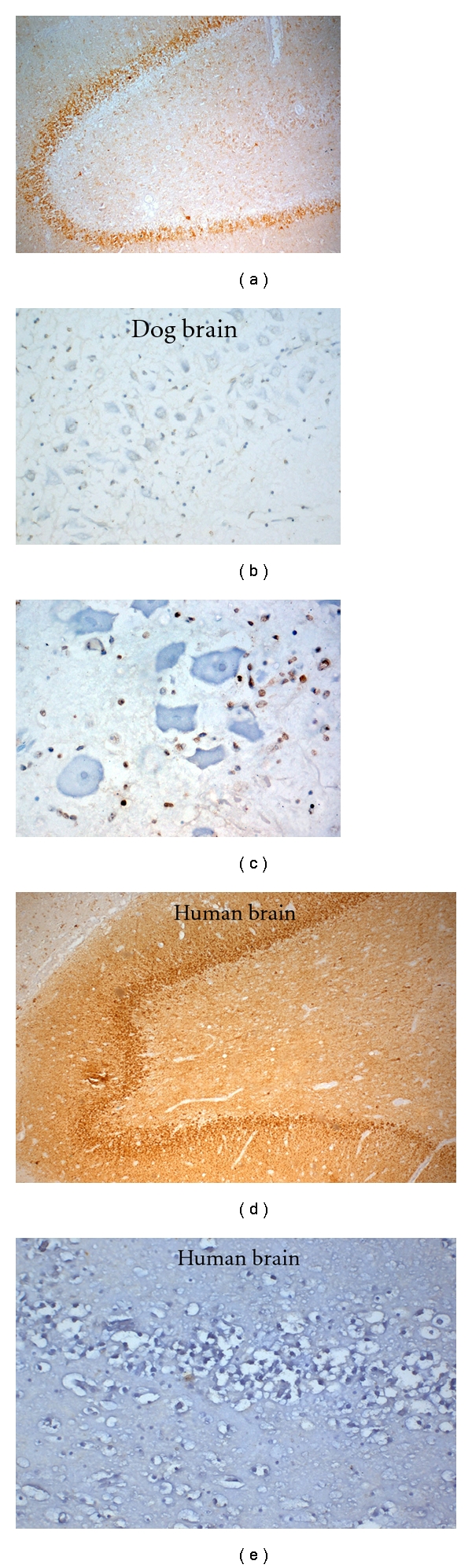
To evaluate the role of apoptosis in rabies encephalitis in humans and canines infected with wild-type street virus, in comparison with rodent model infected with street and laboratory passaged CVS strain, we studied postmortem brain tissue from nine humans, six canines infected with street rabies virus, and Swiss albino mice inoculated intramuscularly (IM) and intracerebrally (IC) with street and CVS strains. Encephalitis and high rabies antigen load were prominent in canine and human brains compared to rodents inoculated with street virus. Neuronal apoptosis was detectable only in sucking mice inoculated with CVS strain and minimal in street virus inoculated mice. In a time point study in suckling mice, DNA laddering was noted only terminally (7 days p.i.) following IC inoculation with CVS strain but not with street virus. In weanling and adult mice, apoptosis was restricted to inflammatory cells and absent in neurons similar to human and canine rabies-infected brains. Absence of neuronal apoptosis in wild-type rabies may facilitate intraneuronal survival and replication while apoptosis in inflammatory cells prevents elimination of the virus by abrogation of host inflammatory response.


Figures

Figure 1
Figure 1
Human rabies viral encephalitis, 15 years/male, incubation period: 6 mon. Section from medulla oblongata showing dense perivascular cuffing of lymphomononuclear cells (a). Multiple Negri bodies are seen within granule neurons of hippocampal dentate gyrus (b). Inset shows multiple Negri bodies in hippocampal pyramidal neurons (b, inset, arrow). Anterior horn cells in the cervical segment of spinal cord are surrounded by microglial cells (c) Immunoreactive to CD68 (arrow, d). Note prominent nucleolus in the neuron reflecting viability. (a): HE ×120; (b): HE ×360; (b inset): HE ×360; C: HE ×300; D: Immunoperoxidase, CD68 ×300.
Figure 2
Figure 2
DNA laddering in Mouse brain. (a) DNA laddering (arrows) is seen only in suckling mouse brain infected with CVS strain of rabies virus (lanes 5 & 6). No laddering seen in street-virus-infected suckling mouse brain (lanes 3 & 4) and weanling adult mouse brain infected with street (lane 7) and CVS strain (lane 8) of rabies virus. (b) DNA laddering (arrows) in suckling mouse brain infected with CVS strain of rabies virus (lanes 3 & 8) and absence of laddering in street-virus-infected adult mouse brain infected with CVS and street strain of rabies virus. (c) DNA laddering (arrows) in suckling mouse brain infected with CVS strain of rabies virus and absence of laddering in street-virus-infected dog brain and human brain. (d) DNA laddering (arrows) in suckling mouse brain infected with CVS strain of rabies virus-time point study. DNA laddering exhibited only on the 6th day P.I.
Figure 3
Figure 3
Suckling mouse brain infected with CVS strain of rabies virus shows rabies viral antigen within the pyramidal neurons of hippocampus (a). Infected pyramidal neurons (b) and glial cells (c) reveal apoptosis by TUNEL immunolabeling. Endothelial cells lining vessels are negative. Weanling mouse brain infected intramuscularly with CVS strain of rabies virus (d-e) shows rabies viral antigen pyramidal neurons of hippocampus (d) and absence of TUNEL labeling in these neurons (e). Weanling mouse brain infected intramuscularly with street strain of rabies-virus-labeled neurons in hippocampus (f) and TUNEL labeling is negative (g). Brain from adult mouse infected intramuscularly with street and CVS strain of rabies virus, respectively, labeled the neurons (f, h) but no apoptosis seen by TUNEL technique (g, i).
Figure 4
Figure 4
Intense immunolabeling of hippocampal pyramidal cells in canine brain naturally infected with street rabies virus (a) are negative for apoptosis by TUNEL labeling (b). Few inflammatory cells in nuclear area of medulla oblongata are labeled by TUNEL technique (c). No neuronal apoptosis noted in these anatomical areas. Pyramidal neurons in human hippocampus from a case of paralytic rabies infected by street virus showed viral antigen (d) in the absence of TUNEL labeling (e). (a: Immunoperoxidase x40; b: TUNEL ×160; c: TUNEL ×400; D: Immunoperoxidase ×40; E: TUNEL ×160.)

References

    1. WHO. Expert Consultation on Rabies . Geneva, Switzerland: WHO; 2005. (Technical Report Series 931). - PubMed
    1. Sudarshan MK, Madhusudana SN, Mahendra BJ, et al. Assessing the burden of human rabies in India: results of a national multi-center epidemiological survey. International Journal of Infectious Diseases. 2007;11(1):29–35. - PubMed
    1. Dietschold B, Rupprecht CE, Fu ZF, Koprowski H. Rhabdo virus. In: Fields B, Knipe D, Howley PM, et al., editors. Fields Virology. 3rd edition. Philadelphia, Pa, USA: Lippincott-Raven; 1996. pp. 1137–1159.
    1. Constantine DG. Rabies transmission by nonbite route. Public Health Reports. 1962;77:287–289. - PMC - PubMed
    1. Hemachudha T. Human rabies: clinical aspects, pathogenesis, and potential therapy. Current Topics in Microbiology and Immunology. 1993;187:122–143. - PubMed

LinkOut - more resources