Review
. 2011 Oct;286(3-4):189-213.
doi: 10.1007/s00438-011-0643-y. Epub 2011 Aug 30.

The lens in focus: a comparison of lens development in Drosophila and vertebrates

Affiliations
Review

The lens in focus: a comparison of lens development in Drosophila and vertebrates

Mark Charlton-Perkins et al. Mol Genet Genomics. 2011 Oct.

Abstract

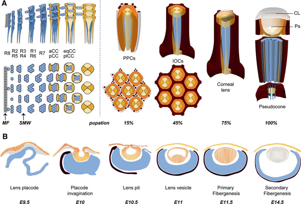
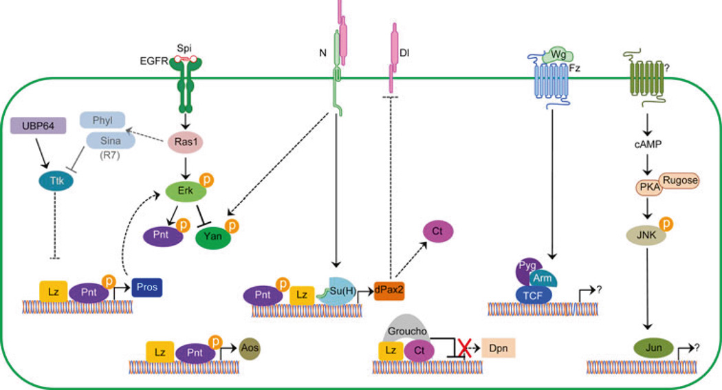
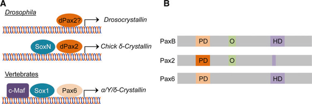
The evolution of the eye has been a major subject of study dating back centuries. The advent of molecular genetics offered the surprising finding that morphologically distinct eyes rely on conserved regulatory gene networks for their formation. While many of these advances often stemmed from studies of the compound eye of the fruit fly, Drosophila melanogaster, and later translated to discoveries in vertebrate systems, studies on vertebrate lens development far outnumber those in Drosophila. This may be largely historical, since Spemann and Mangold's paradigm of tissue induction was discovered in the amphibian lens. Recent studies on lens development in Drosophila have begun to define molecular commonalities with the vertebrate lens. Here, we provide an overview of Drosophila lens development, discussing intrinsic and extrinsic factors controlling lens cell specification and differentiation. We then summarize key morphological and molecular events in vertebrate lens development, emphasizing regulatory factors and networks strongly associated with both systems. Finally, we provide a comparative analysis that highlights areas of research that would help further clarify the degree of conservation between the formation of dioptric systems in invertebrates and vertebrates.


Figures

Fig. 1
Fig. 1
Schematic representation of invertebrate and vertebrate eye development. a In the Drosophila third larval stage, the morphogenetic furrow (MF) moves anteriorly across the eye imaginal disc, blocking proliferative progenitors in G1. From this progenitor pool, the R8, R2/R5, and R3/R4 cells of the neural retina (light blue) are recruited. This is followed by the second mitotic wave, where the remaining progenitors undergo one additional round of mitosis. From these progenitors, the remaining photoreceptors, R1/R6 and R7 are recruited. After photoreceptor recruitment is complete, the anterior and posterior cone cells (aCC/pCC) (yellow) and equatorial and polar cone cells (eqCC/plCC) (light orange) are recruited pairwise. In early pupation, primary pigment cells (PPCs, dark orange) are added, which together with the CCs, complete the full complement of the lens-secreting cells. Soon afterwards, specification of interommatidial pigment cells (dark red) occurs and any remaining non-specified progenitors are eliminated by apoptosis. By ~45% pupation, all cells in the eye begin to terminally differentiate. In the lens, this includes secretion of the corneal lens (CL), followed by secretion of the pseudocone (Ps). IOCs accumulate pigment, while the light-sensing apical membranes (rhabdomeres) of the photoreceptors (PRs) elongate. CCs span the depth of the retina, with the feet ensheathing the PR axons proximally, and the apical surfaces secreting the corneal lens and pseudocone distally. b The onset of lens development in the vertebrate eye is first observed as a thickening of the surface ectoderm into the lens placode (orange) and requires the underlying neural ectoderm (blue) and surrounding mesenchyme (grey). In mouse, this occurs at embryonic day 9.5 (E9.5). The lens placode then invaginates to form the lens pit which pinches off to form the lens vesicle. Primary fibers first form to create a lifelong central core in the lens. Proliferation of precursors continues in the anterior epithelial layer (AEL) (orange nuclei) and the formation and continued addition of secondary fiber cells occurs at the equator and periphery of the lens. The neural retina (light blue) is surrounded by the pigmented epithelium (black). The vitreous humor (white) occupies the space between the lens and retina
Fig. 2
Fig. 2
Schematic representation of cone cell specification. Summary of intrinsic and extrinsic factors involved in the differentiation of the lens-secreting cells in the fly eye. Solid lines are indicative of direct targets while dashed lines represent genetic interactions. Note that Sina and Phyl are expressed in the R7 to prevent Ttk expression, thus preventing R7 photoreceptors from becoming lens-secreting CCs. See text for further details
Fig. 3
Fig. 3
Morphology of wild-type and mutant fly lens facets. Scanning electron microscopy (a) and light microscopy (b) of the external surface of a wild-type adult fly eye shows the characteristic repeating hexagonal arrangement of corneal lens facets. Immunostaining of pupal retinas with E-cad (magenta c, c″) shows the highly regular, organized patterning of the primary pigment cells and interommatidial cells (see Fig. 5 for more details), while the transcripton factor Cut (green c′, c″) reveals the quartet of four cone cells present in the center of each ommatidia. Circles highlight individual ommatidia. d–f In spapol mutants, disorganized, fused, and smaller lens facets are apparent as a rough eye by scanning electron microscopy (d) and a “sparkling” appearance by light microscopy (e). Developmentally, this phenotype results from loss of an average of one CC per ommatidia, aberrant PPC differentiation, and premature IOC death (Fu and Noll 1997; Siddall et al. 2003; Charlton-Perkins et al. 2011), apparent by E-cad (magenta f, f″) and Cut (green f, f″) expression
Fig. 4
Fig. 4
Crystallin regulation by PaxB-derived dPax2 and Pax6. a Currently, only one Crystallin-encoding gene, Drosocrystallin, has been studied in Drosophila. Drosocrystallin expression is lost in dPax2 mutants, but whether dPax2 regulates this gene directly or indirectly is not clear (Dziedzic et al. 2009). Interestingly, however, the chicken δ-Crystallin enhancer, normally regulated by vertebrate Pax6 and Sox1, is activated in Drosophila CCs by dPax2 and SoxN (Blanco et al. 2005), revealing that common regulatory mechanisms are likely to be used to control lens-specific gene expression in vertebrates and invertebrates. b dPax2 and Pax6 appear to be derived from a common ancestor, PaxB, which contains a paired domain (PD) and homeodomain (HD) similar to Pax6, and an octapeptide (O) region that is found in Pax2 (Kozmik et al. 2003). While the PD of dPax2 shares little sequence identity with the PD of Pax6, they are capable of binding similar sequences (Blanco et al. 2005)
Fig. 5
Fig. 5
Expression of N- and E-Cadherin in the Drosophila and mouse lens. a In the fly lens, by mid-pupation, E-cadherin (magenta) is expressed at the apical junction of all cells while N-cad is exclusively expressed at the junctions between the cone cells. a anterior CC, p posterior CC, eq equatorial CC, pl polar CC, PPC primary pigment cell, 2° secondary pigment cell, 3° tertiary pigment cell, B bristle. b In the mouse lens, shown here at embryonic day 11.5, E-Cadherin (magenta) is lost from posterior lens vesicle cell membranes as these cells initiate lens primary fiber cell (FC) differentiation, while N-Cadherin (green) is expressed by all elongated lens fiber cells. The establishment of the anterior epithelial layer (AEL) boundary at the lens equator or transition zone (TZ) is apparent and maintained into adulthood
Fig. 6
Fig. 6
Intrinsic and extrinsic factors used during vertebrate lens development. The intrinsic factors Pax6, Sox2, Foxe3, Pitx1, Sox1, c-Maf, and Prox1 are required at different stages of lens development. Some of the extrinsic factors pathways involved in lens development are summarized here. Fgf signaling: there are 22 fibroblast growth factor ligands (Fgf) ligands and four receptors (Fgfr) in vertebrates, with some variability in ligands among species. Fgfs are secreted morphogens that act both short- and long-range to induce cell proliferation, morphogenesis, and cell type differentiation. Ligands bind to a heterodimeric receptor (Fgfr) complex having a C-terminal tyrosine kinase domain that phosphorylates intracellular proteins upon ligand binding. Targets of Fgfr protein kinase activity include Ras and adaptor proteins Frs, Sos and Grb2 (reviewed in Bottcher and Niehrs 2005). Activated Ras initiates a protein phosphorylation cascade— Ras to Raf to Mek to Erk—which terminates with translocation of phospho-Erk into the nucleus to stimulate factors such as Fos and Ets factors. Er81 and Erm are ETS factors expressed in the lens (Pan et al. 2010). Ras also leads to phosphatidylinositol 3 kinase (PI3K) and AKT activation to promote cell survival during lens fiber differentiation (Weber and Menko 2006). Notch signaling: the Notch pathway transduces a direct, cell-to-cell signal initiated by the binding of a membrane-bound ligand (Jagged or Delta-like proteins) on one cell to a Notch receptor on an adjacent cell (reviewed in Fortini 2009; Kopan and Ilagan 2009). Ligand binding triggers a series of proteolytic cleavages to the Notch receptor, ultimately releasing the Notch intracellular domain (NICD), which translocate into the nucleus where it forms a complex with the transcriptional regulators Rbpj/Su(H) and Mastermind-like (MAML) to activate transcription of target genes such as the bHLH repressors Hes1 or Hes5. The ligand Jag1 is critically required during lens fiber cell differentiation (Le et al. 2009). Bmp/Tgfβ signaling: the transforming growth factor β (Tgfβ) gene superfamily includes the Tgfβ, bone morphogenetic protein (Bmp), growth and differentiation factor (Gdf), Activin and Nodal subfamilies. These secreted ligands bind to heterodimeric Type I and Type II receptor complexes (reviewed in Yang 2004; Moustakas and Heldin 2009). There are seven Type I receptor genes and five Type II receptor genes in vertebrates, all containing cytoplasmic serine/ threonine kinase domains. Upon ligand binding, an active Type II receptor phosphorylates the cytoplasmic domain of a Type I receptor, activating its kinase activity, which then phosphorylates intracellular Smad effectors and other proteins. An activated Smad then complexes with a common mediator, Smad4, and translocates into the nucleus to regulate downstream gene transcription. These signals can be modulated by natural agonists, like Noggin, or by negative feedback from inhibitory Smads (reviewed in Moustakas and Heldin 2009). In lens development, Bmpr1a and Acvr1 are important and signal via Smad5. The Wnt/βcatenin Pathway: Wnts (wingless-type MMTV integration site proteins) are secreted signaling molecules that bind to a Frizzled (Fz) receptor and Lrp co-receptor. Once formed, this complex activates Dishevelled (Dvl), recruits Axin to the cell membrane, and this prevents phosphorylation of the intracellular β-catenin (Ctnnb1). This stabilized β-catenin protein translocates to the nucleus where it interacts with Tcf/Lef transcription factors, as well as the Pygopus (Pyg) co-activator to activate target genes. (reviewed in Fuhrmann 2008). There are 19 Wnt ligands and 10 Fz receptors in vertebrates, of which at least six ligands (Wnts 5a, 5b, 7a, 7b, 8a and 8b), five Fz receptors (Fz 1, 2, 3, 4, 6), two co-receptors (Lrp 5 and 6) and multiple pathway agonists (Dkk, Sfrps) are all expressed in the developing mouse lens (Stump et al. 2003)

Similar articles

Cited by

References

    1. Artavanis-Tsakonas S, Rand MD, Lake RJ. Notch signaling: cell fate control and signal integration in development. Science. 1999;284:770–776. - PubMed
    1. Ashery-Padan R, Marquardt T, Zhou X, Gruss P. Pax6 activity in the lens primordium is required for lens formation and for correct placement of a single retina in the eye. Genes Dev. 2000;14:2701–2711. - PMC - PubMed
    1. Azuma N, Hirakiyama A, Inoue T, Asaka A, Yamada M. Mutations of a human homologue of the Drosophila eyes absent gene (EYA1) detected in patients with congenital cataracts and ocular anterior segment anomalies. Hum Mol Genet. 2000;9:363–366. - PubMed
    1. Bajpe PK, van der Knaap JA, Demmers JA, Bezstarosti K, Bassett A, van Beusekom HM, Travers AA, Verrijzer CP. Deubiquitylating enzyme UBP64 controls cell fate through stabilization of the transcriptional repressor tramtrack. Mol Cell Biol. 2008;28:1606–1615. - PMC - PubMed
    1. Baker NE, Yu SY. The EGF receptor defines domains of cell cycle progression and survival to regulate cell number in the developing Drosophila eye. Cell. 2001;104:699–708. - PubMed

Publication types

MeSH terms

LinkOut - more resources