Review
. 1990 Apr;11(2):82-92.
doi: 10.1097/00003446-199004000-00003.

Outer hair cell electromotility and otoacoustic emissions

Affiliations
Review

Outer hair cell electromotility and otoacoustic emissions

W E Brownell. Ear Hear. 1990 Apr.

Abstract

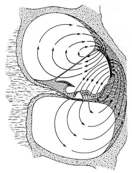
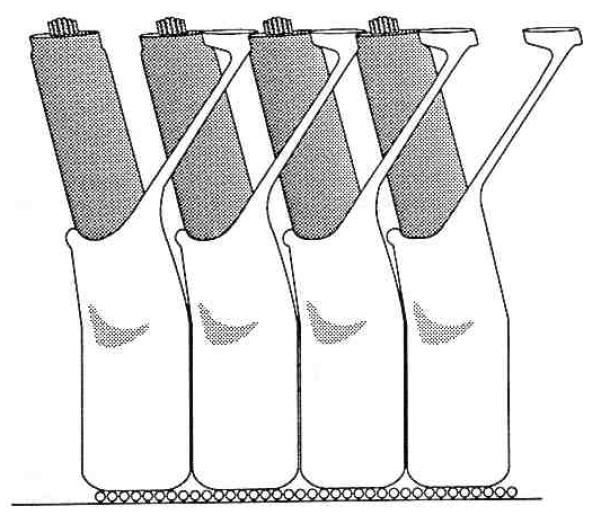
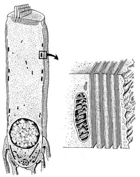
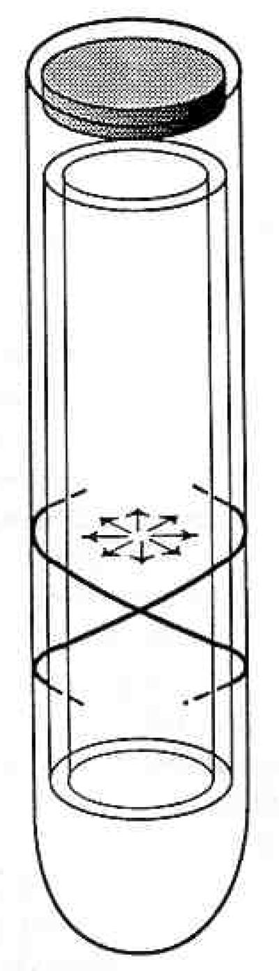
Outer hair cell electromotility is a rapid, force generating, length change in response to electrical stimulation. DC electrical pulses either elongate or shorten the cell and sinusoidal electrical stimulation results in mechanical oscillations at acoustic frequencies. The mechanism underlying outer hair cell electromotility is thought to be the origin of spontaneous otoacoustic emissions. The ability of the cell to change its length requires that it be mechanically flexible. At the same time the structural integrity of the organ of Corti requires that the cell possess considerable compressive rigidity along its major axis. Evolution appears to have arrived at novel solutions to the mechanical requirements imposed on the outer hair cell. Segregation of cytoskeletal elements in specific intracellular domains facilitates the rapid movements. Compressive strength is provided by a unique hydraulic skeleton in which a positive hydrostatic pressure in the cytoplasm stabilizes a flexible elastic cortex with circumferential tensile strength. Cell turgor is required in order that the pressure gradients associated with the electromotile response can be communicated to the ends of the cell. A loss in turgor leads to loss of outer hair cell electromotility. Concentrations of salicylate equivalent to those that abolish spontaneous otoacoustic emissions in patients weaken the outer hair cell's hydraulic skeleton. There is a significant diminution in the electromotile response associated with the loss in cell turgor. Aspirin's effect on outer hair cell electromotility attests to the role of the outer hair cell in generating otoacoustic emissions and demonstrates how their physiology can influence the propagation of otoacoustic emissions.


Figures

Figure 1
Figure 1
Schematic of the electrical environment that powers outer hair cell electromotility (from Zidanic & Brownell, 1990). The standing (or silent) current is represented in terms of current density field lines. The energy from oxidative phosphorylation in the lateral wall of scala media is used to establish an electrochemical gradient across the organ of Corti. The modulation of the silent current results in outer hair cell receptor potentials which are thought to drive rapid electromotility in vivo.
Figure 2
Figure 2
Drawing of a portion of a single row of outer hair cells and associated Deiter’s cells as viewed from the otic capsule looking toward the modiolus. The apical or high frequency end of the cochlea is to the right, lower frequencies are to the left. The three longitudinal domains of the organ of Corti are from top to bottom: (1) the reticular lamina, (2) the fluid space between the reticular lamina, and (3) the Deiter’s cell body layer. The reticular lamina is composed of the apical ends of the hair cells together with the ends of the Deiter’s cells phalangeal processes that interdigitate between them. The Deiter’s cells sit on the basilar membrane (represented by the row of small circles as its constituent fibers project from the plane of the figure).
Figure 3
Figure 3
Outer hair cell morphology. The general organization of the outer hair cell is shown on the left and a blowup of the cell’s cortex is portrayed on the right. The smooth unbroken membranes of the laminated cisternal system are based on the recent description of Evans (1990). Filamentous structures (not portrayed) that may contain actin (Flock, 1988) can be found in the space between the lateral cytoplasmic membrane and the outermost subsurface cistern (see Fig. 4). Stereociliar actin filaments are embedded in the cuticular plate at the top of the cell. Cytoskeletal elements are also found in the intranuclear or synaptic region but not in the central cytoplasmic core of the cell between the cuticular plate and the nucleus.
Figure 4
Figure 4
Schematic of outer hair cell as a pressure vessel. “Circumferential” tensile elements are indicated by oppositely oriented helices in the lateral walls of the cell (Bannister et al, 1988; Flock, 1988; Holley & Ashmore, 1986; Lim et al, 1989). The radially oriented arrows indicate the cytoplasm’s positive hydrostatic pressure. The outer hair cell’s turgor pressure is most likely based on a slightly hyperosmotic cytoplasm that “inflates” the cell.
Figure 5
Figure 5
Pressure gradients in the axial portion of the cytoplasm are required to explain the movement of cytoplasm associated with rapid electromotility. Pressure effects could be transmitted to the cochlear partition even if the cell is isometrically constrained in situ.

Similar articles

Cited by

References

    1. Alberts B, Bray D, Lewis J, Raff M, Roberts K, Watson JD. Molecular Biology of the Cell. Garland; New York: 1983.
    1. Ashmore JF, Brownell WE. Kilohertz movements induced by electrical stimulation in outer hair cells isolated from the guinea pig cochlea. J Physiol. 1986;377:41P.
    1. Ashmore JF, Meech RW. Ionic basis of membrane potential in outer hair cells of guinea pig cochlea. Nature. 1986;322:368–371. - PubMed
    1. Assad JA, Hacohen N, Corey DP. Voltage dependence of adaptation and active bundle movement in bullfrog saccular hair cells. Proc Natl Acad Sci USA. 1989;86:2918–2922. - PMC - PubMed
    1. Bannister LH, Dodson HC, Astbury AF, Douek EE. The cortical lattice: a highly order system of subsurface filaments in guinea pig cochlear outer hair cells. Prog Brain Res. 1988;74:213–219. - PubMed

Publication types