Review
. 2011 Oct;31(10):769-79.
doi: 10.1089/jir.2011.0029. Epub 2011 Aug 30.

The dysregulation of cytokine networks in systemic lupus erythematosus

Affiliations
Review

The dysregulation of cytokine networks in systemic lupus erythematosus

Sokratis A Apostolidis et al. J Interferon Cytokine Res. 2011 Oct.

Abstract

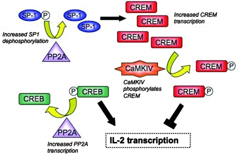
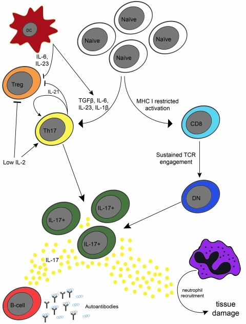
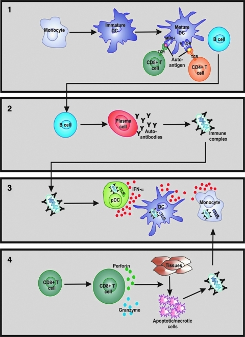
Systemic lupus erythematosus (SLE) is an autoimmune disease associated with chronic immune activation and tissue damage. Organ damage in SLE results from the deposition of immune complexes and the infiltration of activated T cells into susceptible organs. Cytokines are intimately involved in every step of the SLE pathogenesis. Defective immune regulation and uncontrolled lymphocyte activation, as well as increased antigen presenting cell maturation are all influenced by cytokines. Moreover, expansion of local immune responses as well as tissue infiltration by pathogenic cells is instigated by cytokines. In this review, we describe the main cytokine abnormalities reported in SLE and discuss the mechanisms that drive their aberrant production as well as the pathogenic pathways that their presence promotes.


Figures

FIG. 1.
FIG. 1.
Dysregulation of IL2 transcription in SLE T cells. The levels of CREB and CREM dictate IL-2 production by T cells. In healthy individuals, there is a natural balance of these molecules that leads to IL-2 production as needed. In SLE T cells, CREM transcription is enhanced due to increased binding of SP-1 to the CREM promoter. This is facilitated by the dephosphorylation of SP-1 by PP2A, a phosphatase that is found at elevated levels in SLE T cells. SLE, systemic lupus erythematosus; IL, interleukin; CREB, cyclic AMP responsive element-binding protein; CREM, cyclic AMP responsive element modulator.
FIG. 2.
FIG. 2.
The generation and effector functions of IL-17 producing T cells in SLE. TCRαβ double-negative (DN) T cells and CD4+ Th17 T cells comprise the pool of IL-17-producing T cells in SLE. The generation of these 2 subsets is quite different: DN T cells derive from CD8+ T cells after sustained TCR activation, whereas the generation of Th17 T cells is favored in an inflammatory setting, where activated dendritic cells (DCs) provide inflammatory cytokines. This favors Th17 differentiation over production of regulatory T (Treg) cells. The decreased levels of IL-2 in SLE contribute further to the Th17/Treg imbalance. IL-17+ cells infiltrate the tissues, where they induce neutrophil recruitment, causing tissue inflammation, and damage. In addition, IL-17 induces autoantibody formation, by acting on B- cells. TCR, T cell receptor.
FIG. 3.
FIG. 3.
The contribution of IFNα to the disruption of peripheral immune tolerance in SLE. (1). IFNα augments differentiation of monocytes into DCs and from immature DCs to mature, autoantigen-presenting DCs. These DCs present autoantigens to T cells and B cells leading to survival, proliferation, and differentiation of autoreactive lymphocytes. (2). IFNα promotes the differentiation of activated B cells into antibody-secreting plasma cells and subsequent formation of immune complexes (ICs). (3). Through the formation of nucleic acid-containing ICs, a positive feedback loop is formed to enhance TLR7/9 activation and IFNα production in pDCs, myeloid DCs, and monocytes. (4). IFNα enhances autoreactive CD8+ T cell maturation contributing to tissue damage through perforin and granzyme release, thus generating novel autoantigens and further assisting IC-driven production of IFNα. IFN, interferon; TLR, Toll-like receptor; pDC, plasmacytoid dendritic cell.

Similar articles

Cited by

References

    1. Agrawal H. Jacob N. Carreras E. Bajana S. Putterman C. Turner S. Neas B. Mathian A. Koss MN. Stohl W. Kovats S. Jacob CO. Deficiency of type I IFN receptor in lupus-prone New Zealand mixed 2328 mice decreases dendritic cell numbers and activation and protects from disease. J Immunol. 2009;183:6021–6029. - PMC - PubMed
    1. Alcocer-Varela J. Alarcon-Segovia D. Decreased production of and response to interleukin-2 by cultured lymphocytes from patients with systemic lupus erythematosus. J Clin Invest. 1982;69:1388–1392. - PMC - PubMed
    1. Altman A. Theofilopoulos AN. Weiner R. Katz DH. Dixon FJ. Analysis of T cell function in autoimmune murine strains. Defects in production and responsiveness to interleukin 2. J Exp Med. 1981;154:791–808. - PMC - PubMed
    1. Aringer M. Stummvoll GH. Steiner G. Koller M. Steiner CW. Hofler E. Hiesberger H. Smolen JS. Graninger WB. Serum interleukin-15 is elevated in systemic lupus erythematosus. Rheumatology (Oxford) 2001;40:876–881. - PubMed
    1. Awasthi A. Carrier Y. Peron JP. Bettelli E. Kamanaka M. Flavell RA. Kuchroo VK. Oukka M. Weiner HL. A dominant function for interleukin 27 in generating interleukin 10-producing anti-inflammatory T cells. Nat Immunol. 2007;8:1380–1389. - PubMed

Publication types

MeSH terms