Review
. 2011 Aug 30:8:109.
doi: 10.1186/1742-2094-8-109.

Wallerian degeneration: the innate-immune response to traumatic nerve injury

Affiliations
Review

Wallerian degeneration: the innate-immune response to traumatic nerve injury

Shlomo Rotshenker. J Neuroinflammation. .

Abstract

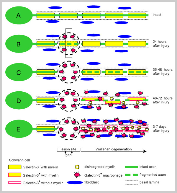
Traumatic injury to peripheral nerves results in the loss of neural functions. Recovery by regeneration depends on the cellular and molecular events of Wallerian degeneration that injury induces distal to the lesion site, the domain through which severed axons regenerate back to their target tissues. Innate-immunity is central to Wallerian degeneration since innate-immune cells, functions and molecules that are produced by immune and non-immune cells are involved. The innate-immune response helps to turn the peripheral nerve tissue into an environment that supports regeneration by removing inhibitory myelin and by upregulating neurotrophic properties. The characteristics of an efficient innate-immune response are rapid onset and conclusion, and the orchestrated interplay between Schwann cells, fibroblasts, macrophages, endothelial cells, and molecules they produce. Wallerian degeneration serves as a prelude for successful repair when these requirements are met. In contrast, functional recovery is poor when injury fails to produce the efficient innate-immune response of Wallerian degeneration.


Figures

Figure 1
Figure 1
Intact and injured PNS nerves. A schematic representation of some of the cellular characteristics of (A) intact and (B through E) injured PNS nerves that undergo normal Wallerian degeneration. (A) Intact myelinating Schwann cells enwrap an intact axon and fibroblasts are scattered between nerve fibers. (B) Traumatic injury produces immediate tissue damage at the lesion site (marked by a circle), a gap (rectangle) may be formed between the proximal and distal nerve stumps, and Galectin-3/MAC-2+ macrophages accumulate at the lesion site within 24 hours after the injury. (C) Destruction of axons is detected during normal Wallerian degeneration 36 hours after the injury. (D) Recruitment of Galectin-3/MAC-2+ macrophages, myelin disintegration, and Galectin-3/MAC-2 expression by Schwann cells begin 48 to 72 hours after injury during normal Wallerian degeneration. (E) Galectin-3/MAC-2+ macrophages and Schwann cells scavenge degenerated myelin during normal Wallerian degeneration, and Schwann cells further proliferate and form Bünger bands.
Figure 2
Figure 2
Intact axon, normal Wallerian degeneration, and kinetics of myelin clearance and Galectin-3/MAC-2 expression during normal Wallerian degeneration. (A) A Schwann cell that is surrounded by basal lamina (arrow heads) forms a myelin sheath around an intact axon; Bar 1 μm. (B) Axons are not detected 7 days after the injury, and Schwann cells (S) and a macrophage (m), which are situated within basal lamina sheaths (dark arrow heads), contain myelin fragments and lipid droplets in their cytoplasm (white arrow heads) (after [16]); Bar 2 μm. (C) Time course of myelin phagocytosis and degradation (Po) and Galectin-3/MAC-2 protein (Gal-3) production. Phagocytosis and degradation of myelin result in the reduction of tissue content of the myelin specific molecule Po. Nerve segments located 5 millimeters distal to lesion sites were removed from wild-type mice at the indicated times and used to determine tissue levels of Po and Gal-3 by ELISA. Those are presented as percentage of their maximal values that are defined 100% (after [60]).
Figure 3
Figure 3
The time course of cytokine protein secretion during normal Wallerian degeneration. Nerve segments located 5 millimeters distal to lesion sites were removed from wild-type mice at the indicated times and used to condition medium with secreted cytokine proteins that were detected and quantified by ELISA. Values are presented as percentage of maximum secretion which is defined 100% (after [60,86]). The secretion of IL-1α is detected within 6 hours after the injury; not shown here since the method of detection was by a bioassay [87].
Figure 4
Figure 4
The cytokine network of Wallerian degeneration. Injury sets in motion the cytokine network of normal Wallerian degeneration. Intact myelinating Schwann cells enwrap intact axons and further express normally the inflammatory cytokines TNFα and IL-1α mRNAs and the TNFα protein. Traumatic injury at a distant site in the far left (not shown) induces the rapid upregulation of TNFα and IL-1α mRNAs expression and proteins production and secretion by Schwann cells within 5 hours. The nature of the signal(s) that are initiated at the injury site, travel down the axon, and then cross over to Schwann cells are not known (?). Concomitantly, Schwann cell derived TNFα and IL-1α induce resident fibroblasts to upregulate the expression of cytokines IL-6 and GM-CSF mRNAs and the production and secretion of their proteins within 2 to 5 hours after the injury. Inflammatory IL-1β mRNA expression and protein production and secretion are induced in Schwann cells with a delay of several hours. The expression of chemokines MCP-1/CCL2 and MIP-1α/CCL3 are upregulated by TNFα, IL-1β and IL-6 as of day 1 after the injury in Schwann cells, and possibly also in fibroblasts and endothelial cells. In turn, circulating monocytes begin their transmigration into the nerve tissue 2 to 3 days after the injury. Fibroblasts begin producing apolipoprotein-E (apo-E) and Schwann cells Galectin-3/MAC-2 (Gal-3) just before the onset of monocyte recruitment. Apolipoprotein-E and Galectin-3/MAC-2 may drive monocyte differentiation towards M2 phenotype macrophage which further produces apolipoprotein-E and Galectin-3/MAC-2. Macrophages efficiently produce IL-10 and IL-6 and much less TNFα, IL-1α, IL-1β. The anti-inflammatory cytokine IL-10, aided by IL-6, down-regulates productions of cytokines. Schwann cells and fibroblasts produce also LIF. Arrows indicate activation and broken lines down-regulation. Not all possible interactions and molecules produced are shown (e.g. autocrine interactions and the role of GM-CSF inhibitor); see text for additional information. The break-down of axons and myelin, and their phagocytosis are not illustrated here; see, however, Figure 1 and Figure 2.

References

    1. Waller A. Experiments on the section of the glossopharyngeal and hypoglossal nerves of the frog, observations of the alterations produced thereby in the structure of their primitive fibers. Phil Transact Royal Soc London. 1850;140:423–429. doi: 10.1098/rstl.1850.0021. - DOI - PubMed
    1. Stoll G, Jander S, Myers RR. Degeneration and regeneration of the peripheral nervous system: from Augustus Waller's observations to neuroinflammation. J Peripher Nerv Syst. 2002;7:13–27. doi: 10.1046/j.1529-8027.2002.02002.x. - DOI - PubMed
    1. Vargas ME, Barres BA. Why is Wallerian degeneration in the CNS so slow? Annu Rev Neurosci. 2007;30:153–179. doi: 10.1146/annurev.neuro.30.051606.094354. - DOI - PubMed
    1. Camara-Lemarroy CR, Guzman-de la Garza F, Fernandez-Garza NE. Molecular Inflammatory Mediators in Peripheral Nerve Degeneration and Regeneration. Neuroimmunomodulation. 2010;17:314–324. doi: 10.1159/000292020. - DOI - PubMed
    1. Coleman MP, Freeman MR. Wallerian degeneration, wld(s), and nmnat. Annu Rev Neurosci. 2010;33:245–267. doi: 10.1146/annurev-neuro-060909-153248. - DOI - PMC - PubMed

Publication types

MeSH terms

LinkOut - more resources