. 2011 Dec;61(8):1379-88.
doi: 10.1016/j.neuropharm.2011.08.024. Epub 2011 Aug 25.

Nicotinic excitatory postsynaptic potentials in hippocampal CA1 interneurons are predominantly mediated by nicotinic receptors that contain α4 and β2 subunits

Affiliations

Nicotinic excitatory postsynaptic potentials in hippocampal CA1 interneurons are predominantly mediated by nicotinic receptors that contain α4 and β2 subunits

Karen A Bell et al. Neuropharmacology. 2011 Dec.

Abstract

In the hippocampus, activation of nicotinic receptors that include α4 and β2 subunits (α4β2*) facilitates memory formation. α4β2* receptors may also play a role in nicotine withdrawal, and their loss may contribute to cognitive decline in aging and Alzheimer's disease (AD). However, little is known about their cellular function in the hippocampus. Therefore, using optogenetics, whole cell patch clamping and voltage-sensitive dye (VSD) imaging, we measured nicotinic excitatory postsynaptic potentials (EPSPs) in hippocampal CA1. In a subpopulation of inhibitory interneurons, release of ACh resulted in slow depolarizations (rise time constant 33.2 ± 6.5 ms, decay time constant 138.6 ± 27.2 ms) mediated by the activation of α4β2* nicotinic receptors. These interneurons had somata and dendrites located in the stratum oriens (SO) and stratum lacunosum-moleculare (SLM). Furthermore, α4β2* nicotinic EPSPs were largest in the SLM. Thus, our data suggest that nicotinic EPSPs in hippocampal CA1 interneurons are predominantly mediated by α4β2* nicotinic receptors and their activation may preferentially affect extrahippocampal inputs in SLM of hippocampal CA1.


Figures

Figure 1
Figure 1
Confocal images of the injection site into the MS/DBB of Chat-cre animals. Scale bar represents 50 μm. Neurons were visualized with a 10 x dry lens (0.3 N.A., voxel dimensions 0.48 × 0.48 × 6.6 μm). Sequential scanning and 4 x line averaging was used to eliminate crosstalk and minimize background noise. A. Image of presumed cholinergic neurons in the medial septum expressing oChIEF-tdTomato. Images were collected using a 561 DPSS laser. B. Image of neurons labeled with anti-Chat antibody (1:200 Millipore AB144P) coupled to Alexa Fluor 633 (Invitrogen), imaged using the red HeNe laser. C. Overlay of images A and B.
Figure 2
Figure 2
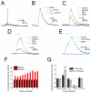
Neurotransmitter release from MS/DBB cholinergic terminals activates α4β2* containing nicotinic receptors on hippocampal CA1 interneurons. A. A blue light flash (blue bar, 1 ms) transiently depolarized a CA1 interneuron. Responses to 5 consecutive stimuli have been superimposed. Two of the 5 stimuli resulted in the production of action potentials. B. Blue light flash produced an excitatory postsynaptic potential (EPSP, black line) in a CA1 interneuron. The EPSP had slow kinetics (green and red dashed lines). C.The EPSP (from B, black) was potentiated by atropine (5 μM, red). Methylycaconitine (MLA, 20 nM, green) did not affect the EPSP. Dihydro-β-erythroidine (DHβE, 10 μM, dark blue) completely inhibited the EPSP. D. A train of blue light flashes (10 × 20 Hz) produced a summating depolarizing plateau response (black) in the same interneuron as B and C. Atropine (5 μM, red) potentiated the depolarization to a greater extent toward later stimuli. MLA (20 nM, green) had no effect on the plateau response. DHβE (10 μM, dark blue) completely inhibited the plateau response. E. In the presence of atropine (5 μM) a train of blue light flashes (10 × 20 Hz) produced a depolarizing response in another interneuron (black). The depolarizing response was unaffected by α-conotoxinPnIA (α-CTX-PnIA, 10 μM, light blue). DHβE (1 μM, dark blue) completely inhibited the depolarizing response. F. Histogram of the effect of atropine (1-5 μM) on the amplitude of the depolarizing nicotinic response produced by a train of blue light flashes (10 × 20 Hz). G.Histogram showing the effect of atropine and nicotinic receptor antagonists on the amplitude of nicotinic EPSPs during a train of blue light flashes (10 × 20 Hz). The amplitudes following the first and tenth flashes have been plotted.
Figure 3
Figure 3
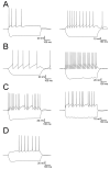
Representative electrophysiological properties of interneurons displaying nicotinic synaptic responses.A. Interneurons with accommodating action potentials (AP) with (left) and without (right) a depolarizing sag during a hyperpolarizing current injection. B. Interneurons with little or no accommodating APs with slow depolarizing sags during hyperpolarizing current pulses. C. Interneurons with irregular or stuttering AP firing patterns with (left) or without (right) depolarizing sags in the membrane potential during hyperpolarizing current pulses. D. An interneuron that displays a delay to AP firing in response to a depolarizing current injection also produced a depolarizing sag in response to hyperpolarizing currents.
Figure 4
Figure 4
Morphology of CA1 interneurons displaying nicotinic EPSPs.Biocytin-labled cells were processed for confocal imaging using strepavidin-633. A. An interneuron with its soma and dendrites localized to the stratum oriens (SO) and axon ramifying in SO and the stratum lacunosum-moleculare (SLM). B. An interneuron with its soma and dendrites localized to the SO and axon ramifying in SO and the stratum radiatum (SR). C. An interneuron with its soma and dendrites primarily localized to the SO and axon projecting toward SLM. D. An interneuron with its soma and dendrites localized primarily to the SLM and some axon in the SLM. E. An interneuron with its soma near the border of SR and SLM, dendrites projecting into the SLM and axon projecting within the expanded stratum pyramidale (SP) and SO. F. An interneuron with its soma in the SLM and dendrites located in all layers of hippocampal CA1.
Figure 5
Figure 5
Synaptic activation of nicotinic receptors primarily occurs on neuronal cell bodies and processes located in SLM. A. Pseudo color images of voltage-sensitive dye (VSD) responses superimposed on the hippocampal slice from which they were measured. Each frame is separated by 25 ms. A burst of blue light flashes (3 × 50 Hz, second frame, blue result of flash artifact) produced depolarizing responses (green – small depolarizations; red – large depolarizations) primarily localized to the SO and SLM. B. Representative VSD responses traces taken from photodiodes overlying each anatomical layer of hippocampal CA1. A burst of blue light flashes produced depolarizing responses in each layer of hippocampal CA1 (black). Atropine (5 μM, red) had little effect on the amplitude of the depolarizing responses. C. Histogram of VSD depolarizing amplitudes (normalized to SLM amplitudes) in each layer of hippocampal CA1 in response to a burst of blue light flashes (black bars). The amplitude of VSD signals in SLM was significantly larger than all other layers of CA1. The VSD signals in SO were significantly larger than those in SR and SP. Atropine (red) did not significantly affect VSD signals in any layer of CA1. D. VSD signals (black) were not inhibited by DNQX (30 μM) and APV (50 μM) (magenta). E. VSD signals in response to a single blue light flashes were unaffected by MLA (50 nM, green) but were inhibited by DHβE (1 μM, dark blue). F. VSD signals in response to a burst of blue light flashes were unaffected by α-CTX-PnIA (10 μM, light blue) but were inhibited by DHβE (5 μM, dark blue). G. Histogram of VSD depolarizing amplitudes normalized to control amplitudes in each layer of hippocampal CA1. DNQX (30 μM) and APV (50 μM) had no effect on VSD amplitudes (magenta bars). MLA (10 to 100 nM) had no significant effect on VSD depolarizing amplitudes (green bars). DHβE (1 to 10 μM) completely inhibited VSD depolarizing amplitudes (dark blue bars). α-CTX PnIA (10 μM) had no effect on VSD depolarizing amplitudes (light blue bar).

References

    1. Alkondon M, Pereira EF, Albuquerque EX. alpha-bungarotoxin- and methyllycaconitine-sensitive nicotinic receptors mediate fast synaptic transmission in interneurons of rat hippocampal slices. Brain Res. 1998;810:257–263. - PubMed
    1. Alkondon M, Pereira EF, Barbosa CT, Albuquerque EX. Neuronal nicotinic acetylcholine receptor activation modulates gamma-aminobutyric acid release from CA1 neurons of rat hippocampal slices. J.Pharmacol.Exp.Ther. 1997;283:1396–1411. - PubMed
    1. Alkondon M, Pereira EF, Eisenberg HM, Albuquerque EX. Choline and selective antagonists identify two subtypes of nicotinic acetylcholine receptors that modulate GABA release from CA1 interneurons in rat hippocampal slices. J.Neurosci. 1999;19:2693–2705. - PMC - PubMed
    1. Atasoy D, Aponte Y, Su HH, Sternson SM. A FLEX switch targets Channelrhodopsin-2 to multiple cell types for imaging and long-range circuit mapping. J.Neurosci. 2008;28:7025–7030. - PMC - PubMed
    1. Atri A, Sherman S, Norman KA, Kirchhoff BA, Nicolas MM, Greicius MD, Cramer SC, Breiter HC, Hasselmo ME, Stern CE. Blockade of central cholinergic receptors impairs new learning and increases proactive interference in a word paired-associate memory task. Behav.Neurosci. 2004;118:223–236. - PubMed

Publication types

MeSH terms