. 2011 Sep 1;124(Pt 17):2976-87.
doi: 10.1242/jcs.085431.

Regulation of sororin by Cdk1-mediated phosphorylation

Affiliations

Regulation of sororin by Cdk1-mediated phosphorylation

Megan R Dreier et al. J Cell Sci. .

Abstract

Tumor cells are commonly aneuploid, a condition contributing to cancer progression and drug resistance. Understanding how chromatids are linked and separated at the appropriate time will help uncover the basis of aneuploidy and will shed light on the behavior of tumor cells. Cohesion of sister chromatids is maintained by the multi-protein complex cohesin, consisting of Smc1, Smc3, Scc1 and Scc3. Sororin associates with the cohesin complex and regulates the segregation of sister chromatids. Sororin is phosphorylated in mitosis; however, the role of this modification is unclear. Here we show that mutation of potential cyclin-dependent kinase 1 (Cdk1) phosphorylation sites leaves sororin stranded on chromosomes and bound to cohesin throughout mitosis. Sororin can be precipitated from cell lysates with DNA-cellulose, and only the hypophosphorylated form of sororin shows this association. These results suggest that phosphorylation of sororin causes its release from chromatin in mitosis. Also, the hypophosphorylated form of sororin increases cohesion between sister chromatids, suggesting that phosphorylation of sororin by Cdk1 influences sister chromatid cohesion. Finally, phosphorylation-deficient sororin can alleviate the mitotic block that occurs upon knockdown of endogenous sororin. This mitotic block is abolished by ZM447439, an Aurora kinase inhibitor, suggesting that prematurely separated sister chromatids activate the spindle assembly checkpoint through an Aurora kinase-dependent pathway.


Figures
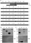
Fig. 1.
Fig. 1.
Cdk1 phosphorylates sororin. (A) Diagram of wild-type and mutant forms of sororin used in this study. Nine potential sites of phosphorylation by Cdk1 are indicated above the wild-type sororin. Each one of these sites appears to be phosphorylated in vivo, as determined by proteomic analysis of phosphopeptides isolated from cells (information obtained from phosida and phosphosite websites) (Beausoleil et al., 2006; Cantin et al., 2008; Chen et al., 2009; Dephoure et al., 2008; Gnad et al., 2007; Olsen et al., 2006; Van Hoof et al., 2009). Each of these nine sites has a serine or threonine followed by proline (minimal Cdk consensus), whereas sites marked with an asterisk conform to the full Cdk consensus ([S/T]Px[K/R]). RDLEM is the potential CIM. Below the wild-type sororin are the various truncation and multi-site mutants of sororin that were generated. In addition, a single mutant at each of the nine sites was generated (not shown). (B) Phosphorylation of sororin by Cdk1 in vitro. Recombinant Cdk1–cyclin B1 was mixed with GST–sororin and a phosphorylation reaction was carried out in vitro with [32P]ATP. Reactions were separated by SDS-PAGE and analyzed by autoradiography. (C) Effect of serine to alanine mutations on sororin phosphorylation. Recombinant sororinWT or sororin9A with an N-terminal GST tag was phosphorylated in an in vitro reaction with Cdk1–cyclin B1 and [32P]ATP. Reactions were analyzed by autoradiography with histone H1 serving as a positive control. CBB, Coomassie-Brilliant-Blue-stained gels.
Fig. 2.
Fig. 2.
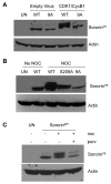
Electrophoretic mobility of sororin. HeLa M cells were transiently transfected with sororinWT–V5 or mutant forms of sororin and analyzed by western blot to detect a mobility shift. (A) Overexpression of Cdk1–cyclin induces a sororin mobility shift. HeLa M cells were blocked in S phase with hydroxyurea and infected with recombinant adenovirus that expressed Cdk1 T14A Y15F (Cdk1-AF) and cyclin B1 fused to the nuclear targeting signal of SV40 T antigen (NB1). Cell lysates were analyzed by western blotting with an antibody to the V5 tag on sororin. (B) Effect of mutations on the electrophoretic migration of sororin. HeLa M cells were transiently transfected with sororin–V5 constructs expressing the indicated proteins. Transfected cells were either left untreated or treated with nocodazole (noc) for 20 hours to block them in mitosis. Cell lysates were separated on a 12.6% SDS-polyacrylamide gel and analyzed by western blotting with an antibody to the V5 tag. (C) Effect of purvalanol A (purv) on the phosphorylation of sororin. Purvalanol A inhibits phosphorylation of sororin. HeLa M cells were transiently transfected with sororinWT-V5, treated with nocodazole with and without purvalanol A for 16 hours, and analyzed by western blotting.
Fig. 3.
Fig. 3.
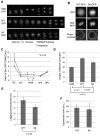
Phosphorylation of sororin releases it from chromatin during mitosis. (A) Localization of sororin–GFP during mitosis. H2B–GFP, sororinWT–GFP and sororin9A–GFP were transiently transfected into HeLa M cells. Time-lapse images of cells progressing through mitosis as seen by fluorescence microscopy (image interval: 12 minutes). Entry into mitosis was indicated by release of sororin–GFP from the nucleus (nuclear envelope breakdown; NEBD). The end of mitosis was indicated by anaphase separation of chromatids. As a control, cells were transiently transfected with H2B–GFP to visualize DNA. In this case, entry into mitosis was taken as the first frame in which DNA condensation was visible. (B) Sororin9A–GFP localizes to DNA in mitosis. Histone H2A–RFP and sororin9A–GFP were transiently transfected into HeLa M cells. An example of a cell showing colocalization of sororin9A–GFP and H2A–RFP is shown. (C) Enrichment of sororinGFP on chromatin. Frames from time-lapse microscopy were used to quantify mean pixel intensities in a defined region of the chromatin and compared with cytoplasmic intensity of the same-sized region. Because cells spent different amounts of time in metaphase, only the first two metaphase frames were quantified. Also, the onset of anaphase occurred at a different time for each cell; this effect is indicated by the dashed line. Averages of at least eight cells with standard errors are shown. (D) SororinWT–V5 and sororin9A–V5 increase the length of mitosis. HeLa M cells were transiently transfected with either sororinWT–GFP or sororin9A–GFP. Frames from time-lapse microscopy were used to quantify the length of mitosis for each cell. (E) Quantification of lagging chromosomes. HeLa M cells were transfected with either sororinWT–V5 or sororin9A–V5, fixed and analyzed by immunofluorescence with antibodies to the V5 tag and to H2A phosphorylated at T121 to indicate centromeres. V5-positive cells in metaphase were assessed for the presence of chromosomes that had not aligned at the metaphase plate. Values are average percentages of cells with lagging chromosomes; bars indicate ± s.e.m. (F) Average expression level of either sororinWT–GFP or sororin9A–GFP in live cells. HeLa M cells were transfected with GFP-tagged sororin and visualized by time-lapse fluorescence microscopy as in A. Single frames of GFP-positive cells in metaphase were used to measure average pixel intensities within the whole cell (a.u., arbitrary units). Values are average from at least 28 cells for each condition; bars indicate ± s.e.m.
Fig. 4.
Fig. 4.
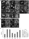
Multiple sites of phosphorylation contribute to the release of sororin from chromatin during prometaphase. SororinWT–GFP and mutant forms of sororin were transiently transfected into HeLa M cells. Transfected cells were exposed to nocodazole to block them in prometaphase. (A) Localization of single point mutants of sororin. Representative cells transfected with the indicated single point mutants of sororin are shown. SororinT159A–GFP showed undetectable levels of fluorescence. (B) Localization of sororin–GFP with multiple S/T to A mutations in HeLa M cells. Specific mutations in each of the constructs are shown in Fig. 1. (C) Staining uniformity of various forms of sororin. To measure the uniformity of staining, digital images were captured of live transfected cells. The standard deviation (SD) of pixel intensities was then determined for each cell. This value was then corrected for the average pixel intensity of each corresponding cell. This corrected standard deviation was averaged over many cells and is shown with standard errors indicated by bars. The various mutants were compared with either sororinWT or sororin9A using a Student's t-test. P-values are indicated in the table.
Fig. 5.
Fig. 5.
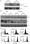
Phosphorylation of sororin reduces its association with the cohesin complex. SororinWT or sororin9A, which contained C-terminal V5 epitope tags, were transiently transfected into HeLa M cells. Some samples were untransfected (UNT). The cells were then synchronized in S phase with 2 mM thymidine for 24 hours. Thymidine was removed and nocodazole was added for 14 hours to synchronize the cells in mitosis. To prepare an S–G2 population, cells were released from the thymidine block for 6 hours. Chromatin was prepared as described in the Materials and Methods. (A) Sororin9A associates with chromatin in mitosis. Cohesin was immunoprecipitated with an antibody to SMC3. Immune complexes were loaded onto 12.6% SDS-polyacrylamide gels and analyzed by western blotting with an anti-V5 antibody. (B) Sororin does not alter the levels of SMC3. Samples were prepared as in A and loaded onto 12.6% SDS-polyacrylamide gels and analyzed by western blotting with SMC3 and anti-V5 antibodies. (C) Verification of the cell cycle stage for each of the cell populations. Flow cytometry was performed using propidium iodide-stained cells. Numbers of cells in G1, S or G2–M were determined from flow cytometry. M, the mitotic index of parallel cultures prepared by the chromosome dropping method; AYSN, asynchronous; UNT, untransfected.
Fig. 6.
Fig. 6.
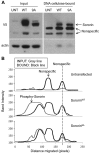
The nonphosphorylated form of sororin binds to DNA–cellulose. (A) Association of sororin with DNA–cellulose. HeLa M cells were transiently transfected with either sororinWT–V5 or sororin9A–V5. UNT, untransfected. Cells were blocked in mitosis by exposure to nocodazole for 16 hours. Cell lysates were incubated with DNA–cellulose for 16 hours and the bound fraction was washed extensively. Proteins remaining associated with the DNA–cellulose were analyzed by western blotting. An aliquot of the lysate used for the binding reaction (Input) was also analyzed for comparison. (B) Desitometric scans of the lanes. The fastest migrating nonspecific band was used to register the scans.
Fig. 7.
Fig. 7.
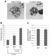
Sororin9A alters sister chromatid cohesion. (A) Examples of chromosome spreads with closed or open arms. SororinWT–V5 and sororin9A–V5 were transfected into HeLa M cells. The cells were then treated for 24 hours with 2 mM thymidine, after which the thymidine was washed off and 100 ng/ml of nocodazole was added for 24 hours. Then chromosome drops were performed. Images of Giemsa-stained cells are shown. Arrows indicate sister chromatids. (B) Quantification of closed sister chromatids in prometaphase. Cells were prepared as in A, and the percentage of spreads where sister chromatids were closed was assessed. Values are averages ± s.e.m. (C) Measurement of interkinetochore distance. HeLa M cells were transfected with sororinWT–V5 or sororin9A–V5 and analyzed by immunofluorescence using antibodies to Hec1 and H2A phosphorylated at T121 [H2A T121(P)]. Antibodies to the V5 tag were also used to identify cells expressing the transfected sororin proteins. H2A T121(P) staining indicates which Hec1-positive dots belong to sister chromatids (see supplementary material Fig. S3B). At least 10 Hec1 pairs per cell were analyzed and at least 14 cells were measured for each condition. Values are averages ± s.e.m.
Fig. 8.
Fig. 8.
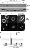
SororinWT–V5 and sororin9A–V5 alleviate a mitotic arrest induced by sororin knockdown. (A) Efficiency of shRNA knockdown of sororin. HeLa M cells were transiently transfected with either a plasmid that produced an shRNA targeting the 3′UTR of sororin (shRNA sororin) or empty pSUPER. Cell lysates were separated on 12.6% SDS-polyacrylamide gels and analyzed by western blotting with an antibody to endogenous sororin. Actin served as a loading control. (B) Defective metaphase plates after sororin knockdown. pSUPER, shRNA sororin or shRNA sororin with sororinWT–V5 were transiently transfected into HeLa M cells. Sororin cDNA clones used in this study lacked a 3′UTR and are not targeted by the shRNA construct. H2B–GFP was also transfected to visualize DNA. Examples of live mitotic cells are shown. (C) Rescue of mitotic arrest by reconstituted sororin. HeLa M cells were transfected simultaneously with a mixture of three plasmids: (1) shRNA against sororin in pSUPER; (2) either sororinWT–V5 or sororin9A–V5 in a mammalian expression construct; and (3) H2B–GFP to visualize the DNA and to mark transfected cells. Positively transfected cells were then quantified 3 days later to determine whether they were in mitosis or interphase. Multinucleate cells were also counted. Living cells were quantified to avoid loss of mitotic cells during fixation.
Fig. 9.
Fig. 9.
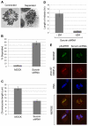
Knockdown of sororin causes activation of the spindle assembly checkpoint. (A) Giemsa-staining of chromosomes to show connected and separated chromosomes. The ‘connected’ spread was from mock-transfected HeLa M cells, and the ‘separated’ spread was from HeLa M cells transfected with sororin shRNA. (B) Chromatid separation after sororin knockdown. HeLa M cells transfected with pBABEpuro (mock) or sororin shRNA were analyzed by chromosome dropping 72 hours post-transfection. (C) Sororin shRNA decreases chromosome length. Cells were transfected as in B and chromosome spreads were analyzed using Slidebook software to measure chromosome lengths. (D) Effect of ZM447439 on mitotic arrest induced by sororin knockdown. HeLa M cells were co-transfected with H2B–GFP, used to mark transfected cells, and with sororin shRNA. 72 hours later, the length of mitosis of GFP-positive cells were determined by time-lapse microscopy. In the +ZM sample, 2.5 μM ZM447439 was added 72 hours post-transfection and filming began at the time of drug addition. Values are averages ± s.e.m. (E) Phosphorylated CENP-A in sororin knockdown cells. HeLa M cells were transfected with sororin shRNA, and 72 hours later analyzed by immunofluorescence microscopy. Cells were stained with antibodies to INCENP and CENP-A phosphorylated at Ser7. Examples of mitotic cells are shown.

Similar articles

Cited by

References

    1. Beausoleil S. A., Villen J., Gerber S. A., Rush J., Gygi S. P. (2006). A probability-based approach for high-throughput protein phosphorylation analysis and site localization. Nat. Biotechnol. 24, 1285-1292 - PubMed
    1. Cantin G. T., Yi W., Lu B., Park S. K., Xu T., Lee J. D., Yates J. R., 3rd (2008). Combining protein-based IMAC, peptide-based IMAC, and MudPIT for efficient phosphoproteomic analysis. J. Proteome Res. 7, 1346-1351 - PubMed
    1. Chen R. Q., Yang Q. K., Lu B. W., Yi W., Cantin G., Chen Y. L., Fearns C., Yates J. R., 3rd, Lee J. D. (2009). CDC25B mediates rapamycin-induced oncogenic responses in cancer cells. Cancer Res. 69, 2663-2668 - PMC - PubMed
    1. Dephoure N., Zhou C., Villen J., Beausoleil S. A., Bakalarski C. E., Elledge S. J., Gygi S. P. (2008). A quantitative atlas of mitotic phosphorylation. Proc. Natl. Acad. Sci. USA 105, 10762-10767 - PMC - PubMed
    1. Diaz-Martinez L. A., Gimenez-Abian J. F., Clarke D. J. (2007). Regulation of centromeric cohesion by sororin independently of the APC/C. Cell Cycle 6, 714-724 - PubMed

Publication types

Substances