. 2011 Sep 6;108(36):14789-94.
doi: 10.1073/pnas.1017825108. Epub 2011 Aug 30.

Improved vascular organization enhances functional integration of engineered skeletal muscle grafts

Affiliations

Improved vascular organization enhances functional integration of engineered skeletal muscle grafts

Jacob Koffler et al. Proc Natl Acad Sci U S A. .

Erratum in

  • Proc Natl Acad Sci U S A. 2012 Jan 24;109(4):1353. Yulia, Shandalov [corrected to Shandalov, Yulia]; Dana, Egozi [corrected to Egozi, Dana]; Daria, Amiad Pavlov [corrected to Pavlov, Daria Amiad]

Abstract

Severe traumatic events such as burns, and cancer therapy, often involve a significant loss of tissue, requiring surgical reconstruction by means of autologous muscle flaps. The scant availability of quality vascularized flaps and donor site morbidity often limit their use. Engineered vascularized grafts provide an alternative for this need. This work describes a first-time analysis, of the degree of in vitro vascularization and tissue organization, required to enhance the pace and efficacy of vascularized muscle graft integration in vivo. While one-day in vitro was sufficient for graft integration, a three-week culturing period, yielding semiorganized vessel structures and muscle fibers, significantly improved grafting efficacy. Implanted vessel networks were gradually replaced by host vessels, coupled with enhanced perfusion and capillary density. Upregulation of key graft angiogenic factors suggest its active role in promoting the angiogenic response. Transition from satellite cells to mature fibers was indicated by increased gene expression, increased capillary to fiber ratio, and similar morphology to normal muscle. We suggest a "relay" approach in which extended in vitro incubation, enabling the formation of a more structured vascular bed, allows for graft-host angiogenic collaboration that promotes anastomosis and vascular integration. The enhanced angiogenic response supports enhanced muscle regeneration, maturation, and integration.


Conflict of interest statement

The authors declare no conflict of interest.

Figures
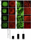
Fig. 1.
Fig. 1.
In vitro grafts organization dynamics. Confocal images of triculture grafts seeded for 1 d, 1 w, 2 w, or 3 w. Green: HUVEC-GFP. Red: Desmin. (AB). Visualizing tissue dynamics on the scaffold. ECs and myoblasts spread toward the perimeter within one day of cell seeding. One week later, ECs had anastomosed and created vessel-like structures, while myoblasts fused to create myofibers. Open vessel-like structures were observed along grafts perimeter (white arrow heads). Two and three weeks later, open vessel-like structures were observed at the center of the scaffold as well, demonstrating strengthening of the structures and close resemblance to natural blood vessels network. Myoblasts already populated the scaffold by day one and fused to form long fibers, as incubation times increased. Bar: A-500 μm. B-1 d, 1 w-100 μm. 2 and 3 w: 200 μm. (C). Diameter of anastomosed HUVEC-GFP structures. Results are presented as mean ± SD, n = 3, p < 0.005. Asterisks indicate statistical significance.
Fig. 2.
Fig. 2.
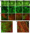
Grafts integration, 14 d postimplantation. (A). Intravital images of the mouse abdomen wall. Grafts are marked with red frame. Grafts were connected to the host through large vessels branching from the epigastric vessels (white arrowheads). Bar-1 mm. (B). Confocal images of grafts. Large host vessels are visible, branching to microvessels upon reaching the graft (white arrowheads). Thin host microvessels (white asterisks) can be seen as they bend and penetrate the graft. These microvessels aligned in parallel after extended incubation times in vitro. Bar-200 μm. (C). Confocal images of fiber organization and integration with vessels. Green—FITC-dextran, red—Desmin. Bar-100 μm. (D). Higher magnification of (C), representing the most advanced integration of fibers and vessels. Bar-100 μm. (E). Confocal image of a normal abdominal muscle showing fibers in red (desmin) and blood vessels in green (FITC-dextran), aligned and parallel. Bar-50 μm. Headings represent in vitro incubation periods.
Fig. 3.
Fig. 3.
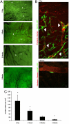
Engineered vessel integration, 14 d postimplantation (A). Intravital images of triculture grafts including HUVEC-GFP followed by systemic perfusion of rhodamin-dextran. ECs differentiated to vessels (white arrowheads) and integrated with host vessels (black). GFP vessels were gradually decreased as in vitro incubation times extended. Bar: 1 d-200 μm, 1 w-100 μm, 2 w-50 μm, 3 week-200 μm. (B). Confocal image of grafts containing HUVEC-GFP perfused with rhodamin-dextran, Functional HUVEC-GFP vessels can be seen, as rho-dextran flows through them. Bar: 1 d-50 μm, 3 w-10 μm. (C). HUVEC-GFP density. ECs gradually disappeared with extended incubation time in vitro. n = 5, p < 0.001. Asterisks indicate statistical significance.
Fig. 4.
Fig. 4.
Vasculogenic and myogenic parameter quantitation, 14 d postimplantation. (A). FITC-dextran signal in the grafts. Perfusion was better in grafts containing engineered blood vessels. Among those, perfusion improved with extended in vitro incubation periods. n = 5, p < 0.001. (B). Total vessel density. Three-week in vitro grafts exhibit the highest number of vessels. n = 5, p < 0.01. (C). Total SMA coverage. Three-week in vitro grafts exhibit the highest vessel coverage. n = 5, p < 0.0001. (D). Mean vessel diameter: Vessel diameters measured on grafts. In vivo vessel diameter increased over the first two weeks of grafts in vitro incubation, followed by a decrease compared to the diameter observed in adjacent normal rectus abdominis muscle. n = 5, p < 0.001. (E). Distribution of vessel diameters: Decrease in diameter was further demonstrated by distribution analysis, where small vessels, under 10 μm in diameter, were observed again in three-week in vitro graft. (F). Capillary density measured by C/F ratio: An overall increase in C/F ratios suggests progressive graft integration. n = 5, p < 0.0005. (G). Mean fiber diameter. n = 5, p < 0.0005. All results are presented as mean ± SD Results in (D) are presented with S.E. Asterisks indicate statistical significance.
Fig. 5.
Fig. 5.
In vivo functionality. (A). Empty constructs showed negligible active stress developed for the different lengths (n = 4), (B). Triculture grafts showed significantly increased active stress (n = 12). (C). Curvilinear force-length relationship could be observed only in triculture grafts. p < 0.05.

References

    1. Atisha D, Alderman AK. A systematic review of abdominal wall function following abdominal flaps for postmastectomy breast reconstruction. Ann Plast Surg. 2009;63:222–230. - PubMed
    1. Baechler MF, Groth AT, Nesti LJ, Martin BD. Soft tissue management of war wounds to the foot and ankle. Foot Ankle Clin. 2010;15:113–138. http://www.ncbi.nlm.nih.gov/pubmed/19067424. - PMC - PubMed
    1. de Vries Reilingh TS, et al. Autologous tissue repair of large abdominal wall defects. Brit J Surg. 2007;94:791–803. - PubMed
    1. Falco EE, Roth JS, Fisher JP. Skeletal muscle tissue engineering approaches to abdominal wall hernia repair. Birth Defects Res C Embryo Today. 2008;84:315–321. http://www.ncbi.nlm.nih.gov/pubmed?term=20189120%5Buid%5D. - PubMed
    1. Mertsching H, et al. Generation and transplantation of an autologous vascularized bioartificial human tissue. Transplantation. 2009;88:203–210. - PubMed

Publication types