. 2011 Oct 14;286(41):36142-36151.
doi: 10.1074/jbc.M111.268847. Epub 2011 Aug 30.

Swapping small ubiquitin-like modifier (SUMO) isoform specificity of SUMO proteases SENP6 and SENP7

Affiliations

Swapping small ubiquitin-like modifier (SUMO) isoform specificity of SUMO proteases SENP6 and SENP7

Kamela O Alegre et al. J Biol Chem. .

Abstract

SUMO proteases can regulate the amounts of SUMO-conjugated proteins in the cell by cleaving off the isopeptidic bond between SUMO and the target protein. Of the six members that constitute the human SENP/ULP protease family, SENP6 and SENP7 are the most divergent members in their conserved catalytic domain. The SENP6 and SENP7 subclass displays a clear proteolytic cleavage preference for SUMO2/3 isoforms. To investigate the structural determinants for such isoform specificity, we have identified a unique sequence insertion in the SENP6 and SENP7 subclass that is essential for their proteolytic activity and that forms a more extensive interface with SUMO during the proteolytic reaction. Furthermore, we have identified a region in the SUMO surface determinant for the SUMO2/3 isoform specificity of SENP6 and SENP7. Double point amino acid mutagenesis on the SUMO surface allows us to swap the specificity of SENP6 and SENP7 between the two SUMO isoforms. Structure-based comparisons combined with biochemical and mutagenesis analysis have revealed Loop 1 insertion in SENP6 and SENP7 as a platform to discriminate between SUMO1 and SUMO2/3 isoforms in this subclass of the SUMO protease family.


Figures
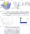
FIGURE 1.
FIGURE 1.
Deconjugation analysis of Loop 1 mutant constructs in SENP7. a and b, superposition of SENP2 and SENP7 catalytic domains in pink and blue, respectively. The position of SUMO2 is indicated schematically based on SUMO2 in complex with SENP2 (Protein Data Bank code 2IO0). SENP7 Loop 1 is indicated in red to highlight the relative position in respect to the entire protease. Key residues are highlighted in stick representation and labeled accordingly. c, sequence alignment of SENP proteases based on structural similarity highlighting Loop 1 insertion in SENP6 and -7. d and g, deconjugation assays of SENP7 and associated mutants at 0.5 nm enzyme concentration against diSUMO2 and polySUMO2, respectively. Assays were run were run at time intervals indicated above each lane in minutes. e, kinetic analysis of deconjugation of diSUMO2 by SENP7 and associated mutants taken at 0, 15, 30, 60, and 120 min. f, bar representation of approximate initial rate velocities for deconjugation of diSUMO2 determined within a linear range from data obtained from e. Axes are labeled, and error bars were obtained by conducting assays in triplicate.
FIGURE 2.
FIGURE 2.
Deconjugation of diSUMO2(D71K) with SENP7 Loop 1 mutant constructs. a, deconjugation assays of SENP7 and associated mutants against diSUMO2(D71K). Assays were run at 5 nm enzyme concentration, and time intervals are indicated above each lane in minutes. b, bar representation of approximate initial rate velocities for deconjugation of diSUMO2(D71K) determined within a linear range based on data obtained from kinetic analysis taken at 0, 15, 30, 60, and 120 min (not shown). Axes are labeled, and error bars were obtained by conducting assays in triplicate.
FIGURE 3.
FIGURE 3.
SENP7-SUMO interaction models, processing and deconjugation activites of SUMO isoform mutants. a, structural model of potential interaction sites between SENP7 (blue ribbon) and SUMO1 (gray ribbon) and SUMO2 (yellow ribbon). The positions of SUMO1 and SUMO2 are based on SUMO1 and -2 in complex with SENP2 (Protein Data Bank codes 1THZ and 2IO0). Key residues in the SUMO-protease interface are indicated in stick representation and labeled according to their position and side chain composition in both SUMO and SENP7. b, electrostatic potential surface representation of SUMO1 and -2 to highlight differences within SENP7 Loop-1 possible interaction sites. All images were modified and represented using the respective Protein Data Bank codes in PyMOL (36). c and d, activity and time course assays, respectively, of deconjugation of SENP2, SENP6, and SENP7 against RanGAP1-SUMO1 (RG-Su), RanGAP1-SUMO1(A72N/H75D), RanGAP1-SUMO2, and RanGAP1-SUMO2(N68A/D71H). An asterisk indicates RanGAP1-SUMO degradation products. e, time course assay of deconjugation of SENP2, -6, and -7 against diSUMO2 (diSu2) and diSUMO2(N68A/D71H) (diSu2m). f, activity assay of processing of preSUMO1GGGiGi-X, preSUMO1GGGiGi-X (A72N/H75D), preSUMO2GGGiGi-X, and preSUMO2GGGiGi-X(N68A/D71H). Activity assays were run at 0.5, 5, and 50 nm enzyme concentration against 5 μm substrate concentration at 37 °C and stopped after 25 min with SDS loading buffer and analyzed via PAGE. Time course assays were run at 0.5 nm enzyme concentration, and time intervals are indicated above each lane in minutes. Proteins were detected by staining with SYPRO (Bio-Rad).
FIGURE 4.
FIGURE 4.
Kinetic analysis for processing and deconjugation of SENP6 deletion mutants. a, deconjugation reaction of RanGAP-SUMO1, RanGAP1-SUMO1(A72N/H75D), RanGAP1-SUMO2, and RanGAP1-SUMO2(N68A/D71H) by SENP6-ΔLoop 1, -ΔLoop 2, and -ΔLoop 3. Activity assays were run at 0.5, 5, and 50 nm enzyme concentration against 5 μm substrate concentration at 37 °C and stopped after 25 min with SDS loading buffer and analyzed via PAGE. Proteins were detected by staining with SYPRO (Bio-Rad). b and c, deconjugation and processing activity, respectively, of SENP6-ΔLoop 3, were taken at 0, 10, 20, 40, and 80 min; deconjugation and processing initial rate activity of SENP6-ΔLoop 3 were taken at 0, 10, 15, and 20 min; bar representation of initial rate velocities for deconjugation and processing of SENP6-ΔLoop 3 were determined within a linear range from data obtained from b. Axes are labeled, and error bars were obtained by conducting assays in triplicate.
FIGURE 5.
FIGURE 5.
Steady-state kinetics of the deconjugation reaction for RanGAP1-SUMO1 and RanGAP1-SUMO1mut by SENP6. a, Deconjugation activity against RanGAP1-SUMO1 and RanGAP1-SUMO1(A72N/H75D) (RG-SUMO1m) with SENP6 and SENP2 at 25 and 1 nm concentrations, respectively. Both substrates are labeled with Alexa Fluor 488, and the kinetics was followed with a VersaDoc apparatus (Bio-Rad). b, Michaelis-Menten graphic representation of substrate concentration (μm) versus velocity (μm min−1) at time intervals 0, 5, 15, 40, and 80 min for RanGAP1-SUMO1mut (left) and RanGAP1-SUMO1 (middle). The right panel depicts a comparison of the two graphs. Initial rate deconjugation activities were measured at five different substrate concentrations (0, 0.25, 1, 2, 6, 20, 50, and 100 μm) and at 25 nm SENP6 concentration. Reactions were stopped after intervals indicated above each lane in minutes with SDS loading buffer and analyzed by gel electrophoresis. c, table of the kinetic coefficients Km, kcat, and kcat/Km obtained from data in b for RanGAP1-SUMO1 and RanGAP1-SUMO1mut.

References

    1. Hershko A., Ciechanover A. (1998) Ann. Rev. Biochem. 67, 425–479 - PubMed
    1. Kerscher O., Felberbaum R., Hochstrasser M. (2006) Annu. Rev. Cell Dev. Biol. 22, 159–180 - PubMed
    1. Geiss-Friedlander R., Melchior F. (2007) Nat. Rev. Mol. Cell Biol. 8, 947–956 - PubMed
    1. Johnson E. S. (2004) Annu. Rev. Biochem. 73, 355–382 - PubMed
    1. Mukhopadhyay D., Dasso M. (2007) Trends Biochem. Sci. 32, 286–295 - PubMed

Publication types

MeSH terms