. 2011 Oct 21;286(42):36509-21.
doi: 10.1074/jbc.M111.268540. Epub 2011 Aug 30.

Group X secreted phospholipase A2 proenzyme is matured by a furin-like proprotein convertase and releases arachidonic acid inside of human HEK293 cells

Affiliations

Group X secreted phospholipase A2 proenzyme is matured by a furin-like proprotein convertase and releases arachidonic acid inside of human HEK293 cells

Ikram Jemel et al. J Biol Chem. .

Abstract

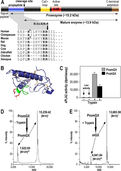
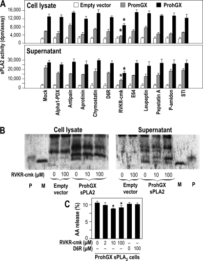
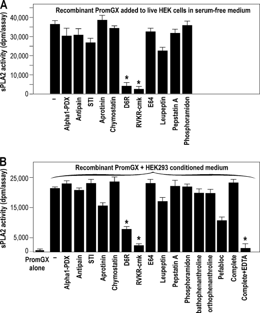
Among mammalian secreted phospholipases A(2) (sPLA(2)s), group X sPLA(2) has the most potent hydrolyzing activity toward phosphatidylcholine and is involved in arachidonic acid (AA) release. Group X sPLA(2) is produced as a proenzyme and contains a short propeptide of 11 amino acids ending with a dibasic motif, suggesting cleavage by proprotein convertases. Although the removal of this propeptide is clearly required for enzymatic activity, the cellular location and the protease(s) involved in proenzyme conversion are unknown. Here we have analyzed the maturation of group X sPLA(2) in HEK293 cells, which have been extensively used to analyze sPLA(2)-induced AA release. Using recombinant mouse (PromGX) and human (ProhGX) proenzymes; HEK293 cells transfected with cDNAs coding for full-length ProhGX, PromGX, and propeptide mutants; and various permeable and non-permeable sPLA(2) inhibitors and protease inhibitors, we demonstrate that group X sPLA(2) is mainly converted intracellularly and releases AA before externalization from the cell. Most strikingly, the exogenous proenzyme does not elicit AA release, whereas the transfected proenzyme does elicit AA release in a way insensitive to non-permeable sPLA(2) inhibitors. In transfected cells, a permeable proprotein convertase inhibitor, but not a non-permeable one, prevents group X sPLA(2) maturation and partially blocks AA release. Mutations at the dibasic motif of the propeptide indicate that the last basic residue is required and sufficient for efficient maturation and AA release. All together, these results argue for the intracellular maturation of group X proenzyme in HEK293 cells by a furin-like proprotein convertase, leading to intracellular release of AA during secretion.


Figures

FIGURE 1.
FIGURE 1.
Structure of group X sPLA2 proenzyme and recombinant production of PromGX and ProhGX sPLA2s. A, schematic representation of group X sPLA2 proenzyme and sequence of the propeptide in various vertebrate species. The likely consensus cleavage site among species RXn(K/R)R is shown. *, position of the Factor Xa miscleavage site within the propeptide that generates miPromGX and miProhGX sPLA2s (the calculated molecular masses of miPromGX and miProhGX are 14,626 and 14,546 Da, respectively). B, three-dimensional model of ProhGX in a schematic representation (blue). Disulfide bridges are highlighted (wheat). The propeptide is displayed in green, and the RR dipeptide side chains are shown in red. The catalytic histidine and the calcium ion are displayed in purple. The picture has been produced using PyMOL. C–E, in vitro activation of PromGX and ProhGX sPLA2s by trypsin. Enzymatic activity was measured using E. coli membranes (C) and MALDI-TOF mass spectrometry analysis (D and E; data only shown for PromGX) before and after treatment of recombinant proenzymes (5 μg) with purified trypsin (50 ng) for 24 h at 37 °C as described under “Experimental Procedures.” Error bars show the S.D. value based on at least duplicate independent analyses. The calculated molecular masses of PromGX and mature mGX are 15,239 and 13,882 Da, respectively. The measured molecular mass values shown are singly (M + H)+ and doubly (M + 2H)2+ charged ions for intact PromGX protein (no trypsin added; D) and for PromGX protein converted into mGX by trypsin (+ trypsin; E).
FIGURE 2.
FIGURE 2.
[3H]-AA release from HEK293 cells treated with exogenous sPLA2s. A, [3H]AA release to the medium from non-transfected HEK293 cells treated with hGX or ProhGX sPLA2s for 6 h as described under “Experimental Procedures.” [3H]AA release is expressed as the percentage of tritium in the culture medium divided by total tritium (medium + cells). B, sPLA2 inhibitor structures used in C. RO-081806A and RO-092906A are hGX sPLA2-specific inhibitors, and RO-032107A is a hGIIA sPLA2-specific inhibitor. C, [3H]AA release to the medium from non-transfected HEK293 cells treated with exogenous hGX sPLA2 and the indicated concentrations of hGX sPLA2-specific inhibitors RO-081806A and RO-092906A (RO08 and RO09, respectively) and the hGIIA sPLA2-specific inhibitor RO-032107A (RO03). Error bars in A and C show the S.D. value based on triplicate independent analyses. *, p < 0.05 versus control (non-stimulated cells, one-way ANOVA with Bonferroni adjustment). #, p < 0.05 versus 200 ng/ml ProhGX sPLA2 (Student's t test).
FIGURE 3.
FIGURE 3.
[3H]AA release from HEK293 cells stably expressing ProhGX and mature hGX (SphGIIA-hGX) sPLA2s. A, [3H]AA release to the medium over 6 h from three different clones of HEK293 cells transfected with empty vector (control), 10 different clones of HEK293 cells transfected with mature hGX sPLA2 (SphGIIA-hGX; i.e. hGX mature sequence fused to the signal peptide of hGIIA sPLA2), and 10 different clones of HEK293 cells transfected with ProhGX sPLA2. B, sPLA2 enzymatic activity in the culture medium of HEK293 cells transfected with empty vector (average of three clones), mature hGX sPLA2 (average of 10 clones), or ProhGX sPLA2 (average of 10 clones). C, [3H]AA release to the medium from transfected HEK293 cells treated with sPLA2 inhibitors at the indicated concentrations. Error bars show the S.D. value based on triplicate independent analyses. *, p < 0.05 versus control (no addition of inhibitor, one-way ANOVA with Bonferroni adjustment). D, [3H]AA release to the medium from HEK293 cells transfected with empty vector, wild-type PromGX sPLA2 (WT), and the PromGX sPLA2 mutants (AA, KA, and AR). Error bars show the S.D. value based on at least duplicate independent analyses. *, p < 0.05 versus empty vector-transfected cells (one-way ANOVA with Bonferroni adjustment). #, p < 0.05 versus WT PromGX sPLA2 (one-way ANOVA with Bonferroni adjustment). E, schematic drawing of the different propeptide constructs of group X sPLA2. The WT sequence and point mutations at the dibasic peptide are indicated. The SphGIIA-hGX construct has no propeptide sequence, and the signal peptide of full-length hGX has been replaced by that of hGIIA sPLA2 (see “Experimental Procedures”).
FIGURE 4.
FIGURE 4.
Processing of PromGX and ProhGX sPLA2s in transfected HEK293 cells. A, sPLA2s were immunoprecipitated with anti-group X sPLA2 antiserum, and the pellet fraction was submitted to Western blot analysis using purified anti-group X sPLA2 IgGs. P and M, recombinant ProhGX and mature hGX sPLA2 standard, respectively. HEK293 cells were transfected with empty vector or mature hGX sPLA2 (SphGIIA-hGX) or ProhGX sPLA2 (two clones shown). The cell supernatant (S) and cell lysate (C) were analyzed. B, the gel shows cells transfected with ProhGX sPLA2 with the cell lysate treated (+G) or not (−G) with N-glycosidase F prior to SDS-PAGE. The Glyco arrowhead marks the position of glycosylated hGX sPLA2. P and M, are ProhGX and mature hGX sPLA2, respectively. C, HEK293 cells expressing propeptide mutants of PromGX sPLA2. WT, cells expressing wild-type PromGX sPLA2; AA, KA, and AR, cells expressing PromGX sPLA2 mutated at the KR dibasic motif of the propeptide. The cell lysate and culture medium cells were analyzed as indicated. P and M, PromGX and mature mGX sPLA2 recombinant standard, respectively. *, a nonspecific band also seen with the empty vector. D, levels of sPLA2 enzymatic activity measured using radiolabeled E. coli membranes as substrate in cell supernatant and cell lysate of PromGX HEK293 cells shown in C. E, total amount of mGX protein measured by mGX-specific time-resolved fluoroimmunoassay in cell supernatant and cell lysate of PromGX HEK293 cells shown in C. Data are representative of at least two independent experiments. Error bars, S.D.
FIGURE 5.
FIGURE 5.
Effect of protease inhibitors on processing of PromGX and ProhGX in transfected HEK293 cells. A, effect of protease inhibitors on sPLA2 activity of HEK293 cells expressing PromGX and ProhGX sPLA2s. HEK293 transfectants expressing PromGX or ProhGX sPLA2s or empty vector were grown to subconfluence in 24-well plates, switched to serum-free medium, and treated with different protease inhibitors for 24 h. The inhibitor concentrations were as follows: α1-antitrypsin Portland (Alpha1-PDX), 1 nm; antipain, 50 μg/ml; aprotinin, 10 μg/ml; chymostatin, 30 μg/ml; D6R and RVKR-cmk, 25 μm; E64, 3 μg/ml; leupeptin, 10 μg/ml; pepstatin A, 5 μg/ml; phosphoramidon (P-amidon), 20 μg/ml; soybean trypsin inhibitor (STI), 10 μg/ml. Cell lysates and cell supernatants were prepared and assayed for sPLA2 enzymatic activity using radiolabeled E. coli membranes. *, p < 0.05 versus control (no addition of inhibitor, one-way ANOVA with Bonferroni adjustment). B, Western blot analysis showing the effect of the cell-permeable proprotein convertase inhibitor RVKR-cmk on the processing of ProhGX sPLA2. HEK293 cells transfected with empty vector or ProhGX sPLA2 were treated with the protease inhibitor for 24 h, after which cell lysates and cell supernatants were analyzed by immunoprecipitation and Western blotting as in Fig. 4A. P and M, recombinant ProhGX sPLA2 and mature hGX sPLA2 standard, respectively. C, release of [3H]AA to the culture medium from ProhGX sPLA2-transfected HEK293 cells in the presence or absence of the cell-permeable inhibitor RVKR-cmk or the cell-impermeable inhibitor D6R. *, p < 0.05 versus control (no addition of inhibitor, one-way ANOVA with Bonferroni adjustment). Data are representative of at least two independent experiments. Error bars, S.D.
FIGURE 6.
FIGURE 6.
Effect of protease inhibitors on processing of exogenous recombinant PromGX and ProhGX sPLA2s by non-transfected HEK293 cells in serum-free medium. A, processing of exogenously added recombinant PromGX sPLA2 in live cells in the absence and presence of protease inhibitors. 80% confluent HEK293 cells were incubated in serum-free DMEM for 24 h at 37 °C in the presence of 500 ng of recombinant PromGX sPLA2 and different protease inhibitors as in Fig. 5A. The medium was then collected and analyzed for sPLA2 enzymatic activity using radiolabeled E. coli membranes. Under the sPLA2 assay conditions used, no enzymatic activity was detected for PromGX sPLA2 incubated in the absence of cells and in conditioned medium from cells incubated without recombinant PromGX sPLA2 (data not shown). B, processing of recombinant PromGX sPLA2 by a concentrated conditioned medium (i.e. in the absence of live cells) obtained from HEK293 cells grown in serum-free medium and effect of protease inhibitors. Recombinant PromGX (500 ng) was incubated in 100 μl of Factor Xa buffer with concentrated conditioned medium (3 μl) obtained without serum in the presence or absence of protease inhibitors overnight at 37 °C, and sPLA2 enzymatic activity was measured. The inhibitor concentrations were as described in Fig. 5A. In this case, we could also test the effects of bathophenantroline (1 mm), orthophenanthroline (1 mm), pefabloc (0.5 mg/ml), and the complete Roche Applied Science mixture inhibitor set with and without EDTA (1× according to the manufacturer's instructions). Data are representative of at least three independent experiments. *, p < 0.05 versus control (no addition of inhibitor, one-way ANOVA with Bonferroni adjustment). Error bars, S.D.

References

    1. Schaloske R. H., Dennis E. A. (2006) Biochim. Biophys. Acta 1761, 1246–1259 - PubMed
    1. Lambeau G., Gelb M. H. (2008) Annu. Rev. Biochem. 77, 495–520 - PubMed
    1. Singer A. G., Ghomashchi F., Le Calvez C., Bollinger J., Bezzine S., Rouault M., Sadilek M., Nguyen E., Lazdunski M., Lambeau G., Gelb M. H. (2002) J. Biol. Chem. 277, 48535–48549 - PubMed
    1. Murakami M., Taketomi Y., Girard C., Yamamoto K., Lambeau G. (2010) Biochimie 92, 561–582 - PubMed
    1. Funk C. D. (2001) Science 294, 1871–1875 - PubMed

Publication types

LinkOut - more resources