. 2011 Nov;19(11):2040-7.
doi: 10.1038/mt.2011.174. Epub 2011 Aug 30.

A double-modulation strategy in cancer treatment with a chemotherapeutic agent and siRNA

Affiliations

A double-modulation strategy in cancer treatment with a chemotherapeutic agent and siRNA

Kazuya Nakamura et al. Mol Ther. 2011 Nov.

Abstract

5-Fluorouracil (5-FU) is broadly considered the drug of choice for treating human colorectal cancer (CRC). However, 5-FU resistance, mainly caused by the overexpression of antiapoptotic proteins such as Bcl-2, often leads ultimately to treatment failure. We here investigated the effect of Bcl-2 gene silencing, using small interfering RNA (siRNA) (siBcl-2), on the efficacy of 5-FU in CRC. Transfection of siBcl-2 by a Lipofectamine2000/siRNA lipoplex effectively downregulated Bcl-2 expression in the DLD-1 cell line (a CRC), resulting in significant cell growth inhibition in vitro upon treatment with 5-FU. For in vivo treatments, S-1, an oral formulation of Tegafur (TF), a prodrug of 5-FU, was used to mimic 5-FU infusion. The combined treatment of polyethylene glycol (PEG)-coated siBcl-2-lipoplex and S-1 showed superior tumor growth suppression in a DLD-1 xenograft model, compared to each single treatment. Surprisingly, daily S-1 treatment enhanced the accumulation of PEG-coated siBcl-2-lipoplex in tumor tissue. We propose a novel double modulation strategy in cancer treatment, in which chemotherapy enhances intratumoral siRNA delivery and the delivered siRNA enhances the chemosensitivity of tumors. Combination of siRNA-containing nanocarriers with chemotherapy may compensate for the limited delivery of siRNA to tumor tissue. In addition, such modulation strategy may be considered a promising therapeutic approach to successfully managing 5-FU-resistant tumors.


Figures

Figure 1
Figure 1
Examination of levels of Bcl-2 and Bax protein expression in DLD-1 cells after transfection with siRNA against Bcl-2 in vitro. (a) Western blot analysis on DLD-1 cells after transfection with siBcl-2 or siCont by Lipofectamine 2000 lipoplexes. Bands of Bcl-2 (26 kDa), Bax (20 kDa), and β-actin (42 kDa) were recorded by LAS-4000 EPUVmini. (b) Quantitative evaluation of the percent change in expression levels of Bcl-2 protein against β-actin one. Data represent mean ± SD from three independent experiments. (c) Quantitative evaluation of the percent change in levels of Bax protein against β-actin one. Data represent mean ± SD from three independent experiments. siRNA, small interfering RNA.
Figure 2
Figure 2
Effect of the combined treatment of siBcl2 and 5-FU on viability and apoptosis of DLD-1 cells in vitro. (a) Effect of different concentrations of 5-FU on cell viability of nontreated, siCont-treated, and siBcl-2-treated DLD-1 cells. Cell viability was determined by MTT assay. (b) Effect of single or combined treatment with siBcl-2 (6.25 nmol/l) and 5-FU (0.5 µg/ml) on apoptosis in DLD-1 cells. Apoptotic cells were detected by TUNEL assay. Data were represented from three independent experiments. ***P < 0.001 versus nontreated cells (none). *P < 0.05 siBcl-2 and siBcl-2 + 5-FU. 5-FU, 5-Fluorouracil; MTT, 3-(4,5-dimethylthiazol-2-yl)-2,5-diphenyltetrazolium bromide.
Figure 3
Figure 3
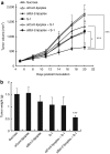
Tumor growth suppressive effect of the combined treatment of PEG-coated siBcl-2-lipoplexes and S-1 in the DLD-1 bearing mouse model. (a) Tumor volume following treatments. Tumor xenografts were established by subcutaneous implantation of DLD-1 cells in nude mice. S-1 (6.9 mg/kg) was orally administered daily from day 5 to 19 after tumor cell inoculation. PEG-coated siRNA-lipoplexes containing either siCont or siBcl2 (10 µg siRNA/mouse) were intravenously administered every 2 days (on day 5, 7, 9, 11, 13, 15, 17, and 19 after tumor cell inoculation). For the control group, sucrose was administered instead of S-1 and PEG-coated siRNA lipoplexes. (b) Tumor weight on day 21 post-tumor inoculation. Data represent mean ± SD (n = 6). ***P < 0.001 versus control. PEG, polyethylene glycol.
Figure 4
Figure 4
Suppression of Bcl-2 protein expression and induction of apoptosis in the tumor tissue after in vivo treatment. (a) Bcl-2 and Bax protein expression was determined by western blot analysis. β-actin protein was used for equal loading assessment. (b) Numbers of apoptotic cells in the tumor section were determined by TUNEL staining. (c) Percent of TUNEL-positive cells in the section. Data represent mean ± SD. ***P < 0.001 versus sucrose. ###P < 0.001 versus S-1 or siBcl-2-lipoplex.
Figure 5
Figure 5
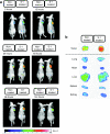
Effect of S-1 treatment on in vivo tumor accumulation of PEG-coated siRNA-lipoplexes. DLD-1 bearing mice were pretreated with or without daily S-1 dosing (6.9 mg tegafur/kg) for 7 days. On the last day of S-1 treatment, mice received an intravenous injection of DiD labeled-PEG-coated siRNA-lipoplexes. One representative picture of three independent experiments was shown here. (a) In vivo imaging of the lipoplexes at 6, 12, 24, 48, 96, and 144 hours postinjection. (b) Organ distribution of the lipoplexes 24 hours postinjection. PEG, polyethylene glycol.
Figure 6
Figure 6
Effect of S-1 treatment on in vivo tumor accumulation and biodistribution of radio-labeled PEG-coated siRNA-lipoplexes. DLD-1 bearing mice were pretreated with or without daily S-1 dosing (6.9 mg tegafur/kg) for 7 days. On the last day of S-1 treatment, mice received an intravenous injection of 3H-CHE-labeled PEG-coated siRNA-lipoplex. At 24 hours after injection, samples were collected and the radioactivity in blood and major organs was determined. (a) Radioactivity (%dose) in tumor tissues. (b) Radioactivity (%dose) in blood, lung, liver, spleen, and kidney. Data represent mean ± SD (n = 3–4). **P < 0.01 versus control. PEG, polyethylene glycol.

References

    1. Subbarayan PR, Sarkar M, Nelson G, Benitez E, Singhal S., and, Ardalan B. Chronic exposure of colorectal cancer cells in culture to fluoropyrimidine analogs induces thymidylate synthase and suppresses p53. A molecular explanation for the mechanism of 5-FU resistance. Anticancer Res. 2010;30:1149–1156. - PubMed
    1. Noordhuis P, Holwerda U, Van der Wilt CL, Van Groeningen CJ, Smid K, Meijer S.et al. (20045-Fluorouracil incorporation into RNA and DNA in relation to thymidylate synthase inhibition of human colorectal cancers Ann Oncol 151025–1032. - PubMed
    1. Wada H, Nagano H, Yamamoto H, Noda T, Murakami M, Kobayashi S.et al. (2009Combination of interferon-alpha and 5-fluorouracil inhibits endothelial cell growth directly and by regulation of angiogenic factors released by tumor cells BMC Cancer 9361. - PMC - PubMed
    1. Nishimura G, Yanoma S, Mizuno H, Satake K, Taguchi T, Ikeda Y.et al. (2000Therapeutic effect of 1 M tegafur-0.4 M 5-chloro-2, 4-dihydroxypridine-1 M potassium oxonate (S-1) on head and neck squamous carcinoma cells Cancer Lett 1591–7. - PubMed
    1. Koga M, Anegawa E, Yoh J, Tsuyama H, Sakaino H, Iwamoto O.et al. (2010Clinical relevance of thymidylate synthase (TS) activity for S-1-based chemotherapy in squamous cell carcinoma of the oral cavity Br J Oral Maxillofac Surg 4888–93. - PubMed

Publication types

MeSH terms