. 2011 Aug 30:2:452.
doi: 10.1038/ncomms1463.

Blimp1 regulates the transition of neonatal to adult intestinal epithelium

Affiliations

Blimp1 regulates the transition of neonatal to adult intestinal epithelium

Vanesa Muncan et al. Nat Commun. .

Abstract

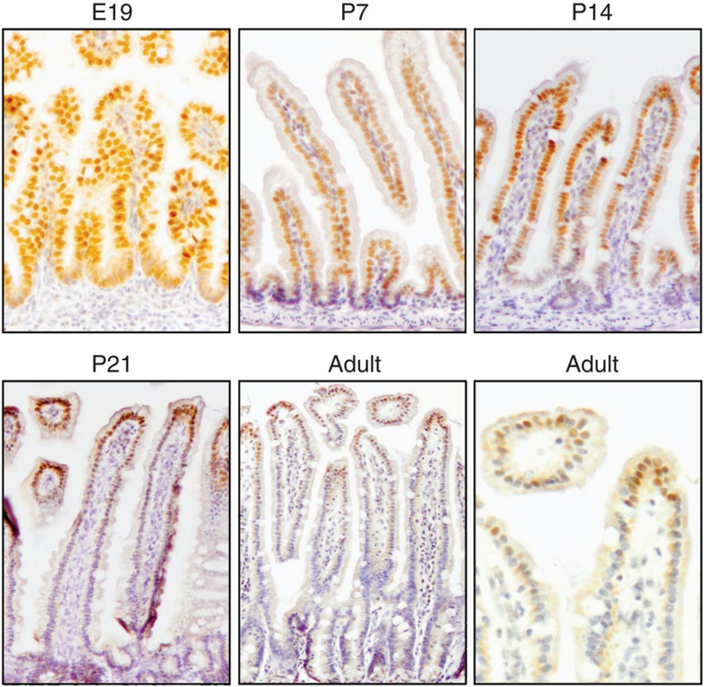
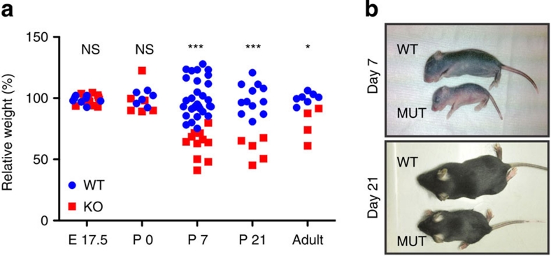
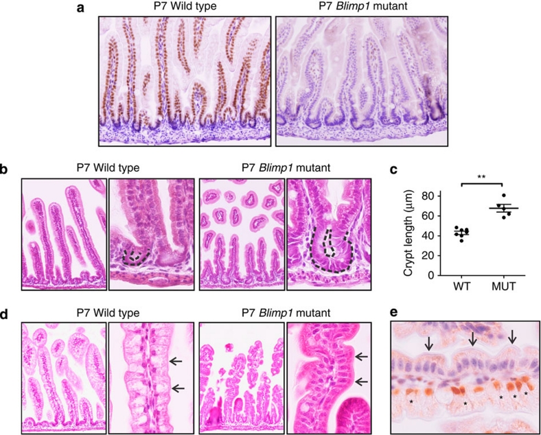
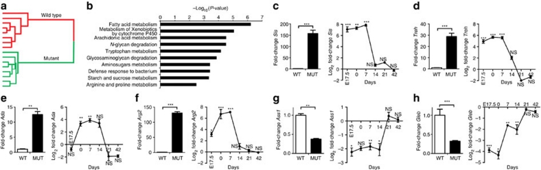
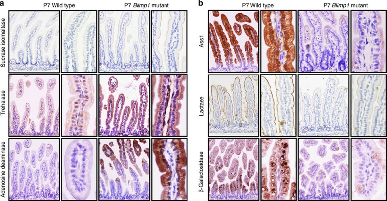
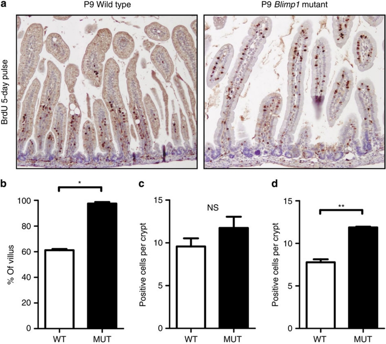
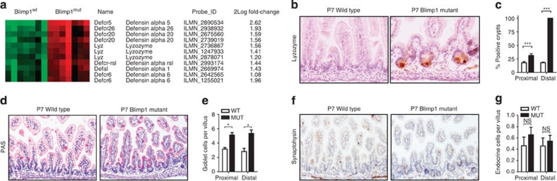
In many mammalian species, the intestinal epithelium undergoes major changes that allow a dietary transition from mother's milk to the adult diet at the end of the suckling period. These complex developmental changes are the result of a genetic programme intrinsic to the gut tube, but its regulators have not been identified. Here we show that transcriptional repressor B lymphocyte-induced maturation protein 1 (Blimp1) is highly expressed in the developing and postnatal intestinal epithelium until the suckling to weaning transition. Intestine-specific deletion of Blimp1 results in growth retardation and excessive neonatal mortality. Mutant mice lack all of the typical epithelial features of the suckling period and are born with features of an adult-like intestine. We conclude that the suckling to weaning transition is regulated by a single transcriptional repressor that delays epithelial maturation.


Figures

Figure 1
Figure 1. Blimp1 is expressed until the suckling to weaning transition in the intestinal epithelium of the mouse.
Immunohistochemistry shows nuclear localization of Blimp1 in the small intestinal epithelium, which becomes restricted to the upper part of the villus around day 21 and finally only marks a few cells at the villus tip in adult mice (arrows in enlargement). Original magnifications: ×200 and ×800 for magnification.
Figure 2
Figure 2. Growth retardation in mice with intestinal epithelial deletion of Blimp1.
(a) Relative weight of Blimp1 mutant and control littermates at different ages. (b) Representative images of Blimp1 mutant and control animals at P7 and P21 shows growth retardation of the mutants. NS, not significant; HET, heterozygote; MUT, mutant; WT, wild type. *P<0.05; ***P<0.001, Student's t-test.
Figure 3
Figure 3. Accelerated maturation of Blimp1 mutant intestinal epithelium.
(a) Immunohistochemistry for Blimp1 shows efficient loss of Blimp1 from the intestinal epithelium. (b) In the proximal small intestine, there was a noticeably enhanced formation of crypts (dotted line) at P7 compared with the rudimentary crypts (dotted line) of the wild-type mice. (c) Average crypt depth in wild type (WT, n=7) and Blimp1 mutant (MUT, n=5) animals, average plus s.e. (d) In the distal small intestine of WT mice, enterocytes clearly showed the translucent cytoplasm that is typical for the presence of large cytoplasmic vacuoles that characterize this stage (arrows). These vacuoles were completely absent in Blimp1 mutant enterocytes (arrows). (e) A rare villus on which Blimp1 is only partially deleted clearly shows the difference in phenotype between Blimp proficient (brown nuclear staining, asterisks) and deficient cells (arrows). **P<0.01, Student's t-test. Original magnifications ×200 for overviews and ×800 for enlargements.
Figure 4
Figure 4. Transmission electron microscopy shows loss of vacuoles and accelerated brush border development in Blimp1 mutants.
(a) Image of the apical canalicular system of a wild-type (WT) mouse. Mutant (MUT) mice completely lack this system. (b) An image of a whole cell clearly shows how the apical canalicular system (ACS) feeds into large cytoplasmic vacuoles (Vs) in WT mice and that this system is absent in Blimp1 mutants. (c) An image of the brush borders of WT and MUT mice shows a much more developed brush border in mutant mice. Bars in left lower corners indicate 1 μm.
Figure 5
Figure 5. A major metabolic shift towards the adult phenotype in Blimp1 mutant (MUT) mice at P7.
(a) Cluster analysis of the individual mice on the gene array showed that the MUT animals group together and are a separate cluster from the wild-type (WT) mice. (b) Most of the pathways that are significantly altered in the MUT mice are metabolic pathways. (ch) Real-time RT–PCR analysis of selected candidates from the significantly altered genes identified in the array. (c) Sucrase isomaltase (Sis), (d) trehalase (Treh), (e) adenosine deaminase (Ada), (f) arginase 2 (Arg2), (g) argininosuccinate synthetase 1 (Ass1), (h) β-galactosidase (Gleb). For each gene the fold difference at P7 is shown on the left and the Log2 fold differences at different time points on the right. For E17.5, P0 and 1, 2, 3 and 6 weeks the number of WT animals is n=5, n=3, n=7, n=5, n=7, n=8, respectively, whereas n=5, n=3, n=8, n=7, n=6 and n=10 for the MUT animals, respectively. *P<0.05; **P<0.01 and ***P<0.001, NS, not significant. Data were analysed using a two-way analysis of variance, mean and s.e. are shown.
Figure 6
Figure 6. Adult-type enzyme expression pattern in Blimp1 mutant (MUT) mice.
Immunohistochemistry shows that (a) the adult-type enzymes sucrase isomaltase, trehalase and adenosine deaminase are aberrantly expressed in MUT mice. (b) In contrast, expression of suckling-period-specific enzymes such as argininosuccinate synthetase 1, lactase and β-galactosidase are lost. Note the localization of β-galactosidase to the cytoplasmic vacuoles in the wild-type animal. Original magnifications ×200 and ×800 for enlargements.
Figure 7
Figure 7. Accelerated intestinal epithelial migration in Blimp1 mutant (MUT) animals.
Animals were injected with BrdU at P4 and killed at P9 to examine the speed of epithelial migration along the crypt–villus axis. (a) Immunohistochemistry for BrdU in wild type (WT) and Blimp1 MUT animals. (b) In WT (n=5) animals, cells had migrated an average of 61% of the villus length compared with 98% in the MUT (n=5) animals. (c) Number of Ki67-positive cells per crypt (n=8 animals per group). (d) Number of BrdU-positive cells per crypt (n=2 animals per group). Average and s.e. value is shown, *P<0.05; **P<0.01, Student's t-test. NS not significant.
Figure 8
Figure 8. Premature development of Paneth cells in Blimp1 mutant (MUT) mice.
(a) Heatmap of specific Paneth cell genes that are upregulated in the mid-intestine of the Blimp1 MUT animals. (b) Immunohistochemistry for lysozyme shows premature presence of Paneth cells in Blimp1 MUT animals at P7. Original magnification ×400. (c) The percentage of crypts containing Paneth cells was larger in MUT mice than in wild types (WTs), with the most remarkable difference in the distal small intestine, ***P<0.001, two-way analysis of variance. (d) Periodic acid-Schiff (PAS) staining shows goblet cells in the distal intestine of WT and Blimp1 MUT mice. (e) Quantification of the number of PAS-positive goblet cells per villus in the proximal (3.2±0.25 versus 5.1±0.34 in WT versus MUT) and distal intestine (2.8±0.38 versus 5.3±0.53 in WT versus MUT). (f) Immunohistochemistry for synaptophysin in WT and Blimp1 MUT mice. (g) Quantification of the number of synaptophysin-positive enteroendocrine cells per villus in the proximal (0.46±0.18 versus 0.67±0.13 in WT versus MUT) and distal intestine (0.46±0.14 versus 0.54±0.10 in WT versus MUT). *P<0.05, two-way analysis of variance. Original magnifications ×200. NS, not significant.

References

    1. Henning S. J. Postnatal development: coordination of feeding, digestion, and metabolism. Am. J. Physiol. 241, G199–G214 (1981). - PubMed
    1. Moxey P. C. & Trier J. S. Development of villus absorptive cells in the human fetal small intestine: a morphological and morphometric study. Anat. Rec. 195, 463–482 (1979). - PubMed
    1. Kraehenbuhl J. P., Bron C. & Sordat B. Transfer of humoral secretory and cellular immunity from mother to offspring. Curr. Top. Pathol. 66, 105–157 (1979). - PubMed
    1. de Santa B. P., van den Brink G. R. & Roberts D. J. Development and differentiation of the intestinal epithelium. Cell Mol. Life Sci. 60, 1322–1332 (2003). - PMC - PubMed
    1. Bry L. et al. Paneth cell differentiation in the developing intestine of normal and transgenic mice. Proc. Natl Acad. Sci. USA 91, 10335–10339 (1994). - PMC - PubMed

Substances