. 2011 Aug 30:2:457.
doi: 10.1038/ncomms1466.

Molecular mechanism for 3:1 subunit stoichiometry of rod cyclic nucleotide-gated ion channels

Affiliations

Molecular mechanism for 3:1 subunit stoichiometry of rod cyclic nucleotide-gated ion channels

Noah G Shuart et al. Nat Commun. .

Abstract

Molecular determinants of ion channel tetramerization are well characterized, but those involved in heteromeric channel assembly are less clearly understood. The heteromeric composition of native channels is often precisely controlled. Cyclic nucleotide-gated (CNG) channels from rod photoreceptors exhibit a 3:1 stoichiometry of CNGA1 and CNGB1 subunits that tunes the channels for their specialized role in phototransduction. Here we show, using electrophysiology, fluorescence, biochemistry, and X-ray crystallography, that the mechanism for this controlled assembly is the formation of a parallel 3-helix coiled-coil domain of the carboxy-terminal leucine zipper region of CNGA1 subunits, constraining the channel to contain three CNGA1 subunits, followed by preferential incorporation of a single CNGB1 subunit. Deletion of the carboxy-terminal leucine zipper domain relaxed the constraint and permitted multiple CNGB1 subunits in the channel. The X-ray crystal structures of the parallel 3-helix coiled-coil domains of CNGA1 and CNGA3 subunits were similar, suggesting that a similar mechanism controls the stoichiometry of cone CNG channels.


Conflict of interest statement

The authors declare no competing financial interests.

Figures

Figure 1
Figure 1. Heteromeric rod CNG channel.
(a) Cartoon of the heteromeric rod CNG channel composed of three CNGA1 subunits (red) and one CNGB1 subunit (green). The intracellular carboxy-terminal domain contains a C-linker, CNBD, and CLZ domain (only on the CNGA subunits). (b) Sequence alignment of CLZ domains of the different CNGA subunits from bovine (b), rat (r) and human (h). Residues predicted to lie in the 'a' and 'd' positions of heptad repeats are shown in magenta.
Figure 2
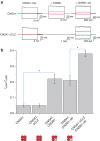
Figure 2. Deletion of the CNGA1 CLZ domain increased the cAMP efficacy of heteromeric CNG channels.
(a) Leak subtracted macroscopic current traces from inside–out patches in the presence of 2.5 mM cGMP (black) and 16 mM cAMP (red) in response to voltage pulses of −60 mV and +60 mV. Currents were recorded for CNGA1 only (top left), CNGA1-ΔCLZ only (bottom left), CNGA1+CNGB1 (top middle), CNGA1-ΔCLZ +CNGB1 (bottom middle), CNGA1+CNGB1-ΔN (top right), CNGA1-ΔCLZ +CNGB1-ΔN (bottom right). (b) IcAMP/IcGMP at +60 mV for each channel composition: CNGA1 (n=6), CNGA1-ΔCLZ (n=6), CNGA1+CNGB1 (n=7), CNGA1-ΔCLZ +CNGB1 (n=7), and CNGA1-ΔCLZ +CNGB1-ΔN (n=10). The expected subunit stoichiometry for each case is shown at the bottom. Data are plotted as mean±s.e.m. Asterisks denote statistically significant differences with P<0.01.
Figure 3
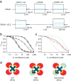
Figure 3. Deletion of the CNGA1 CLZ domain confers intermediate L-cis-diltiazem block on heteromeric CNG channels.
(a) cGMP-activated current traces from inside–out patches in the absence (black) and presence (blue) of 100 μM L-cis-diltiazem in response to voltage pulses of −60 mV and +60 mV. Currents were recorded for CNGA1 only (top left), CNGA1-ΔCLZ only (bottom left), CNGA1+CNGB1 (top middle), CNGA1-ΔCLZ +CNGB1 (bottom middle), CNGA1+CNGB1-ΔN (top right), and CNGA1-ΔCLZ +CNGB1-ΔN (bottom right). (b) Dose-response relations of L-cis-diltiazem block at +60 mV for CNGA1 only (n=5, filled squares), CNGA1-ΔCLZ only (n=6, open squares), CNGA1+CNGB1 (n=5, filled circles), CNGA1+CNGB1-ΔN (n=5, open circles), and CNGA1-ΔCLZ +CNGB1-ΔN (n=4, filled triangles). Data are plotted as mean±s.e.m. Data are fitted with a single Langmuir isotherm or the sum of two Langmuir isotherms (for CNGA1-ΔCLZ +CNGB1-ΔN). The dashed line shows the poor fit of CNGA1-ΔCLZ +CNGB1-ΔN with two Langmuir isotherms with affinities constrained to those of CNGA1-ΔCLZ only and CNGA1+CNGB1-ΔN. (c) Dose-response relation of L-cis-diltiazem block at +60 mV for CNGA1-ΔCLZ +CNGB1-ΔN at an RNA ratio of 1:1 (orange triangles) fitted with the sum of two Langmuir isotherms (orange line). Single Langmuir isotherms from CNGA1 only and CNGA1+CNGB1 are shown in grey and the sum of two Langmuir isotherms from CNGA1-ΔCLZ +CNGB1-ΔN at an RNA ratio of 1:2 is shown in black. (d) Cartoon depicting how interactions between a single molecule of L-cis-diltiazem (blue), and CNGA1 (red) and CNGB1 (green) subunits, could produce an intermediate affinity for channels containing multiple CNGB1 subunits.
Figure 4
Figure 4. FRET experiments reveal multiple CNGB1 subunits when the CNGA1 CLZ domain is deleted.
(a) Predictions for FRET between eCFP-CNGB1-ΔN and eYFP-CNGB1-ΔN for channels containing just a single CNGB1 subunit (left) or possibly multiple subunits (right). Possibilities for FRET are shown by arrows. (b) Emission spectra of Xenopus oocytes expressing CNGA1-ΔCLZ with both eCFP-CNGB1-ΔN and eYFP-CNGB1-ΔN with 458 nm excitation (red) or 488 nm excitation (black). A scaled eCFP only excitation is shown in blue and extracted spectrum (difference between red and blue) is shown in yellow. Ratio A is the ratio of the yellow to the black spectrum. (c) Emission spectra of CNGA1-ΔCLZ+eYFP-CNGB1-ΔN coloured as in (b). Ratio Ao is the ratio of the red to the black spectrum. (d) Ratio A-Ratio Ao for FRET between CNGB1 subunits with (n=8) and without (n=6) the CLZ domain in the CNGA1 subunit. (e) Ratio A-Ratio Ao for FRET between CNGA1 and CNGB1 subunits with (n=6) and without (n=4) the CLZ domain in the CNGA1 subunit. Data are plotted as mean±s.e.m. Asterisks indicate statistically-significant differences, with P<0.05.
Figure 5
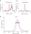
Figure 5. Size-exclusion chromatography and light scattering reveal the CLZ domains from CNGA1 and CNGA3 form trimers.
(a) FSEC results of CNGA1#621–690 (left) and CNGA3#626–694 (right) as N-terminal (red) and C-terminal (black) fusions to GFP. The peaks eluting at 13 mL are the predicted size for a trimer of the fusion proteins. (b) LS-SEC results of CNGA1#621–690 (blue) and CNGA3#626–694 (magenta). The smooth traces show the elution of the protein (ultraviolet absorbance) and the data points show the measured molecular weight (left axis).
Figure 6
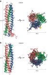
Figure 6. Structure of the trimeric coiled-coil CNGA CLZ complexes.
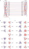
Ribbon representation of the trimeric coiled-coil structures of CNGA1#621–690 (a) and CNGA3#626–672 (b). Each complex is shown from a side (left) and top (right) perspective (top, N-terminus; bottom, C-terminus). The metal ions coordinated by the glutamate and aspartate residues at two sites in CNGA1#621–690 are shown as yellow spheres.
Figure 7
Figure 7. Analysis of the hydrophobic packing in CNGA1 and CNGA3 CLZ domains.
(a) Hydrophobic layers of CNGA1#621–690 (left) and CNGA3#626–672 (right). Side chains are depicted for the 'a' (red) and 'd' (blue) positions of the coiled coils. (b,c) Packing geometry for the 'a' (red) and 'd' (blue) positions of CNGA1#621–690 (b) and CNGA3#626–672 (c).
Figure 8
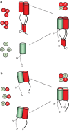
Figure 8. Mechanism for the controlled assembly of heteromeric rod CNG channels.
(a) The CNGA1 subunits (red) initially assemble into trimers through homotypic association of their carboxy-terminal CLZ domains into a parallel three helix coiled coil. Subsequently, a CNGB1 subunit (green), if present, is preferentially incorporated into the remaining slot of the channel tetramer due to a high affinity association with CNGA1 subunits. (b) Deletion of the CLZ domain in CNGA1 allows CNGA1 subunits to assemble with CNGB1 subunits. This produces channels with multiple CNGB1 subunits and a large dominant negative effect of CNGB1.

Similar articles

Cited by

References

    1. Kreusch A, Pfaffinger PJ, Stevens CF, Choe S. Crystal structure of the tetramerization domain of the Shaker potassium channel. Nature. 1998;392:945–948. doi: 10.1038/31978. - DOI - PubMed
    1. Li M, Jan YN, Jan LY. Specification of subunit assembly by the hydrophilic amino-terminal domain of the Shaker potassium channel. Science. 1992;257:1225–1230. doi: 10.1126/science.1519059. - DOI - PubMed
    1. Howard RJ, Clark KA, Holton JM, Minor DL., Jr. Structural insight into KCNQ (Kv7) channel assembly and channelopathy. Neuron. 2007;53:663–675. doi: 10.1016/j.neuron.2007.02.010. - DOI - PMC - PubMed
    1. Tsuruda PR, Julius D, Minor DL., Jr. Coiled coils direct assembly of a cold-activated TRP channel. Neuron. 2006;51:201–212. doi: 10.1016/j.neuron.2006.06.023. - DOI - PMC - PubMed
    1. Jenke M, et al. C-terminal domains implicated in the functional surface expression of potassium channels. EMBO J. 2003;22:395–403. doi: 10.1093/emboj/cdg035. - DOI - PMC - PubMed

MeSH terms

Substances