Review
. 2011 Aug 31;30(17):3501-15.
doi: 10.1038/emboj.2011.266.

Let's go bananas: revisiting the endocytic BAR code

Affiliations
Review

Let's go bananas: revisiting the endocytic BAR code

Britta Qualmann et al. EMBO J. .

Abstract

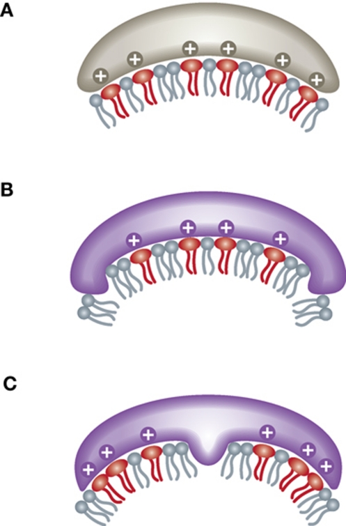
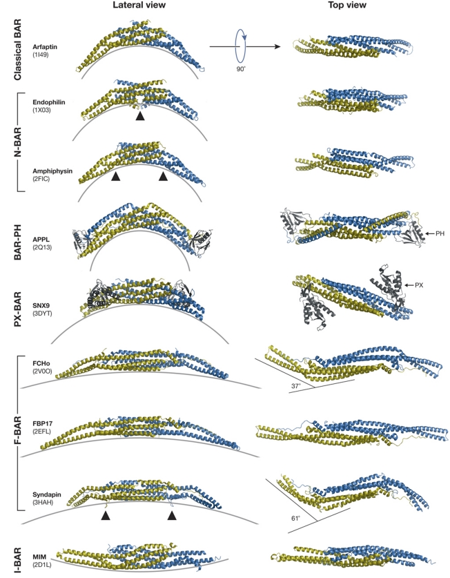
Against the odds of membrane resistance, members of the BIN/Amphiphysin/Rvs (BAR) domain superfamily shape membranes and their activity is indispensable for a plethora of life functions. While crystal structures of different BAR dimers advanced our understanding of membrane shaping by scaffolding and hydrophobic insertion mechanisms considerably, especially life-imaging techniques and loss-of-function studies of clathrin-mediated endocytosis with its gradually increasing curvature show that the initial idea that solely BAR domain curvatures determine their functions is oversimplified. Diagonal placing, lateral lipid-binding modes, additional lipid-binding modules, tilde shapes and formation of macromolecular lattices with different modes of organisation and arrangement increase versatility. A picture emerges, in which BAR domain proteins create macromolecular platforms, that recruit and connect different binding partners and ensure the connection and coordination of the different events during the endocytic process, such as membrane invagination, coat formation, actin nucleation, vesicle size control, fission, detachment and uncoating, in time and space, and may thereby offer mechanistic explanations for how coordination, directionality and effectiveness of a complex process with several steps and key players can be achieved.


Conflict of interest statement

The authors declare that they have no conflict of interest.

Figures

Figure 1
Figure 1
Mechanisms of membrane shaping by BAR domains. (A) Scaffolding mechanism promoted by binding to inverted cone-shaped negatively charged lipids (red). (B, C) Hydrophobic insertion mechanisms embedding protein parts into the lipid layer.
Figure 2
Figure 2
BAR modules adopt folds with different degrees of curvature. BAR modules (one monomer in yellow and the other in blue) from different subfamilies viewed from the side (left) and from top (right). Additional domains (PH and PX) are in grey. PDB codes are given in brackets. Note that FCHo and syndapin have tilde shapes. Arrowheads mark positions within the BAR modules for hydrophobic insertion into membranes.
Figure 3
Figure 3
Vesicle formation correlates with increased curvature. The formation of a clathrin-coated vesicle from pre-existing clathrin-coated plaques (A) and from a clathrin-coated pit (B) is marked by an increase of membrane curvature and a drop of the corresponding circle diameters (black circles in (A) and (B)).
Figure 4
Figure 4
Changes of membrane topology during clathrin-mediated endocytosis. (AC) Detailed view of recruitment traces of BAR superfamily proteins (A), F-BAR proteins (B) and dynamin, F-actin (lifeact) and N-WASP (C). At time t=0, fission of a clathrin-coated vesicle was detected. Images modified from Taylor et al (2011). (D) Colour-coded changes in membrane topology at distinct sites of vesicle formation. (E) Localization of BAR superfamily proteins to different sites of membrane curvature during clathrin-mediated endocytosis. For simplicity, clathrin is omitted. F-BAR domains with very shallow curvature (yellow), shallow curvature (orange) and strongly tilde shape (red) are shown in different orientations of binding. Strongly curved BAR modules are in purple. White bars mark defined positions on the molecules to visualize rotations. (F) Electron micrograph of a stimulated lamprey synapse injected with anti-endophilin antibodies (reprinted from Ringstad et al, 1999 with permission from Elsevier) displays an unusual accumulation of clathrin-coated pits that were arrested at different stages of vesicle formation, that is, with different curvatures (arrows). Bar, 100 nm. Endo2, endophilin A2; Amph1, amphiphysin I; Synd2, syndapin II; Dyn1, dynamin I.

Similar articles

Cited by

References

    1. Andersson F, Jakobsson J, Low P, Shupliakov O, Brodin L (2008) Perturbation of syndapin/PACSIN impairs synaptic vesicle recycling evoked by intense stimulation. J Neurosci 28: 3925–3933 - PMC - PubMed
    1. Andersson F, Low P, Brodin L (2010) Selective perturbation of the BAR domain of endophilin impairs synaptic vesicle endocytosis. Synapse 64: 556–560 - PubMed
    1. Anggono V, Smillie KJ, Graham ME, Valova VA, Cousin MA, Robinson PJ (2006) Syndapin I is the phosphorylation-regulated dynamin I partner in synaptic vesicle endocytosis. Nat Neurosci 9: 752–760 - PMC - PubMed
    1. Aspenström P (1997) A Cdc42 target protein with homology to the non-kinase domain of FER has a potential role in regulating the actin cytoskeleton. Curr Biol 7: 479–487 - PubMed
    1. Ayton GS, Blood PD, Voth GA (2007) Membrane remodeling from N-BAR domain interactions: insights from multi-scale simulation. Biophys J 92: 3595–3602 - PMC - PubMed

Publication types