

PMS: photosystem I electron donor or fluorescence quencher
- PMID: 21879310
- PMCID: PMC3296009
- DOI: 10.1007/s11120-011-9671-z
PMS: photosystem I electron donor or fluorescence quencher
Abstract
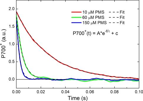
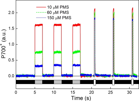
Light energy harvested by the pigments in Photosystem I (PSI) is used for charge separation in the reaction center (RC), after which the positive charge resides on a special chlorophyll dimer called P700. In studies on the PSI trapping kinetics, P700(+) is usually chemically reduced to re-open the RCs. So far, the information available about the reduction rate and possible chlorophyll fluorescence quenching effects of these reducing agents is limited. This information is indispensible to estimate the fraction of open RCs under known experimental conditions. Moreover, it would be important to understand if these reagents have a chlorophyll fluorescence quenching effects to avoid the introduction of exogenous singlet excitation quenching in the measurements. In this study, we investigated the effect of the commonly used reducing agent phenazine methosulfate (PMS) on the RC and fluorescence emission of higher plant PSI-LHCI. We measured the P700(+) reduction rate for different PMS concentrations, and show that we can give a reliable estimation on the fraction of closed RCs based on these rates. The data show that PMS is quenching chlorophyll fluorescence emission. Finally, we determined that the fluorescence quantum yield of PSI with closed RCs is 4% higher than if the RCs are open.
Figures


Similar articles
-
Characterization of the structural changes and photochemical activity of photosystem I under Al(3+) effect.J Photochem Photobiol B. 2015 Aug;149:292-9. doi: 10.1016/j.jphotobiol.2015.06.012. Epub 2015 Jun 15. J Photochem Photobiol B. 2015. PMID: 26123191
-
Structure of the higher plant light harvesting complex I: in vivo characterization and structural interdependence of the Lhca proteins.Biochemistry. 2005 Mar 1;44(8):3065-73. doi: 10.1021/bi047873g. Biochemistry. 2005. PMID: 15723551
-
The Quenching of Long-Wavelength Fluorescence by the Closed Reaction Center in Photosystem I in Thermostichus vulcanus at 77 K.Int J Mol Sci. 2024 Nov 19;25(22):12430. doi: 10.3390/ijms252212430. Int J Mol Sci. 2024. PMID: 39596495 Free PMC article.
-
Light harvesting in photosystem I supercomplexes.Biochemistry. 2006 Jan 17;45(2):331-45. doi: 10.1021/bi051932o. Biochemistry. 2006. PMID: 16401064 Review.
-
Light-harvesting in photosystem I.Photosynth Res. 2013 Oct;116(2-3):153-66. doi: 10.1007/s11120-013-9838-x. Epub 2013 May 4. Photosynth Res. 2013. PMID: 23645376 Free PMC article. Review.
Cited by
-
Photosynthesis: basics, history and modelling.Ann Bot. 2020 Sep 14;126(4):511-537. doi: 10.1093/aob/mcz171. Ann Bot. 2020. PMID: 31641747 Free PMC article. Review.
-
Spectral diversity of photosystem I from flowering plants.Photosynth Res. 2023 Jan;155(1):35-47. doi: 10.1007/s11120-022-00971-2. Epub 2022 Oct 19. Photosynth Res. 2023. PMID: 36260271 Free PMC article.
-
Excitation transfer and trapping kinetics in plant photosystem I probed by two-dimensional electronic spectroscopy.Photosynth Res. 2018 Mar;135(1-3):239-250. doi: 10.1007/s11120-017-0427-2. Epub 2017 Aug 14. Photosynth Res. 2018. PMID: 28808836
-
Light-harvesting complex II is an antenna of photosystem I in dark-adapted plants.Nat Plants. 2020 Jul;6(7):860-868. doi: 10.1038/s41477-020-0693-4. Epub 2020 Jun 22. Nat Plants. 2020. PMID: 32572215
-
Single chloroplast in folio imaging sheds light on photosystem energy redistribution during state transitions.Plant Physiol. 2023 Feb 12;191(2):1186-1198. doi: 10.1093/plphys/kiac561. Plant Physiol. 2023. PMID: 36478277 Free PMC article.
References
-
- Berthold DA, Babcock GT, Yocum CF. A highly resolved, oxygen-evolving photosystem-II preparation from spinach thylakoid membranes—electron-paramagnetic-res and electron-transport properties. FEBS Lett. 1981;134:231–234. doi: 10.1016/0014-5793(81)80608-4. - DOI
Publication types
MeSH terms
Substances
LinkOut - more resources
Full Text Sources
Other Literature Sources
