. 2012 Jan;21(1):40-9.
doi: 10.1007/s00586-011-2004-2. Epub 2011 Aug 31.

Classification of three-dimensional thoracic deformities in adolescent idiopathic scoliosis from a multivariate analysis

Affiliations

Classification of three-dimensional thoracic deformities in adolescent idiopathic scoliosis from a multivariate analysis

Samuel Kadoury et al. Eur Spine J. 2012 Jan.

Abstract

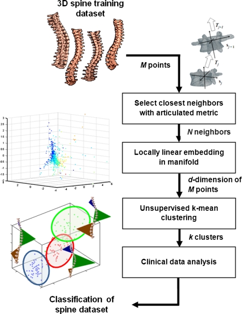
Purpose: Understanding how to classify and quantify three-dimensional (3D) spinal deformities remains an open question in adolescent idiopathic scoliosis. The objective of this study was to perform a 3D manifold characterization of scoliotic spines demonstrating thoracic deformations using a novel geometric and intuitive statistical tool to determine patterns in pathological cases.

Methods: Personalized 3D reconstructions of thoracic (T)/lumbar (L) spines from a cohort of 170 Lenke Type-1 patients were analyzed with a non-linear manifold embedding algorithm in order to reduce the high-dimensionality of the data, using statistical properties of neighbouring spine models. We extracted sub-groups of the data from the underlying manifold structure using an unsupervised clustering algorithm to understand the inherent distribution and determine classes of pathologies which appear from the low-dimensional space.

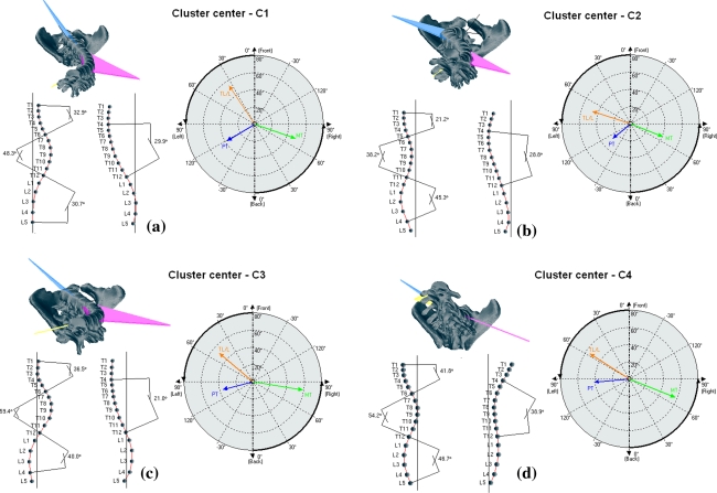
Results: For Lenke Type-1 patients, four clusters were detected from the low-dimensional manifold of 3D models: (1) normal kyphosis (T) with hyper-lordosis (L) and high Cobb angles (37 cases), (2) low kyphosis (T) and normal lordosis (L), with high rotation of plane of maximum curvature (55 cases), (3) hypo-kyphotic (T) and hyper-lordosis (L) (21 cases) and (4) hyper-kyphotic (T) with strong vertebral rotation (57 cases). Results show the manifold representation can potentially be useful for classification of 3D spinal pathologies such as idiopathic scoliosis and serve as a tool for understanding the progression of deformities in longitudinal studies.

Conclusions: Quantitative evaluation illustrates that the complex space of spine variability can be modeled by a low-dimensional manifold and shows the existence of an additional hyper-kyphotic subgroup from the cohort of 3D spine reconstructions of Lenke Type-1 patients when compared with previous findings on the 3D classification of spinal deformities.


Figures

Fig. 1
Fig. 1
Workflow diagram of the proposed method. From a cohort of 3D spine reconstructions, the system sequentially (1) selects the closest neighbors for each patient, (2) embeds the data to a manifold embedding, (3) performs an unsupervised clustering of the data points and (4) analyzes the clusters based on a set of geometrical clinical parameters
Fig. 2
Fig. 2
Illustration of the personalized 3D spine reconstruction from preoperative standing X-rays [18]
Fig. 3
Fig. 3
Anatomical landmark identification on a the thoracic vertebra with endplate centers and pedicle extremity tips and b the pelvis with points on the S1 endplate
Fig. 4
Fig. 4
Graphical representation of 3D clinical indices used for the evaluation of spinal deformities. a Computer Cobb angle in the coronal plane; b Kyphosis and lordosis in the sagital plane; c Axial rotation of the apical vertebra; d Projection of the maximal curvature plane in 3D view; e Plane of maximal curvature in the top view
Fig. 5
Fig. 5
a Evolution of the percentage (%) increase of the number of significant weights with respect to the number of N neighbors. b Evolution of the residual variance with respect to the value of the lower dimension d
Fig. 6
Fig. 6
Result of the low-dimensional manifold of 3D spine models from 170 Lenke Type-1 patients, clustering into four sub-groups. Cluster center points from these groups show (1) normal kyphosis with hyper-lordosis, (2) low kyphosis and normal lordosis, with high rotation of plane of maximum curvature, (3) hypo-kyphotic and hyper-lordosis and (4) hyper-kyphotic cases
Fig. 7
Fig. 7
Frontal, lateral and top view profiles with planes of maximal deformity (PMC) of the cluster centers for the four detected clusters. The respective daVinci representations are also shown with corresponding cases. a Cluster center from C1. b Cluster center from C2. c Cluster center from C3. d Cluster center from C4
Fig. 8
Fig. 8
Trendlines illustrating the distribution of geometrical indices such as the Main Thoracic (MT) Cobb angle, kyphosis, MT plane of maximum curvature and MT apex apical rotation for all 170 patients when analyzing the a 1st dimension, b 2nd dimension and c 3rd dimension of the low-dimensional manifold

References

    1. Clin J, Aubin CE, Parent S, et al. Comparison of the biomechanical 3D efficiency of different brace designs for the treatment of scoliosis using a finite element model. Eur Spine J. 2010;19:1169–1178. doi: 10.1007/s00586-009-1268-2. - DOI - PMC - PubMed
    1. Kadoury S, Cheriet F, Beauséjour M, et al. A three-dimensional retrospective analysis of the evolution of spinal instrumentation for the correction of adolescent idiopathic scoliosis. Eur Spine J. 2009;18:23–37. doi: 10.1007/s00586-008-0817-4. - DOI - PMC - PubMed
    1. Villemure I, Aubin CE, Grimard G et al. (2001) Progression of vertebral and spinal three-dimensional deformities in adolescent idiopathic scoliosis: a longitudinal study. Spine 2226–2244 - PubMed
    1. Kadoury S, Cheriet F, Laporte C, et al. A versatile 3D reconstruction system of the spine and pelvis for clinical assessment of spinal deformities. Med Biol Eng Comput. 2007;45:591–602. doi: 10.1007/s11517-007-0182-1. - DOI - PubMed
    1. Carpineta L, Labelle H. Evidence of three-dimensional variability in scoliotic curves. Clin Orthop Relat Res. 2003;412:139–148. doi: 10.1097/01.blo.0000072462.53786.96. - DOI - PubMed