. 2011 Aug 31:6:10.
doi: 10.1186/1750-2187-6-10.

G-patch domain and KOW motifs-containing protein, GPKOW; a nuclear RNA-binding protein regulated by protein kinase A

Affiliations

G-patch domain and KOW motifs-containing protein, GPKOW; a nuclear RNA-binding protein regulated by protein kinase A

Anne Kristin Aksaas et al. J Mol Signal. .

Abstract

Background: Post-transcriptional processing of pre-mRNA takes place in several steps and requires involvement of a number of RNA-binding proteins. How pre-mRNA processing is regulated is in large enigmatic. The catalytic (C) subunit of protein kinase A (PKA) is a serine/threonine kinase, which regulates numerous cellular processes including pre-mRNA splicing. Despite that a significant fraction of the C subunit is found in splicing factor compartments in the nucleus, there are no indications of a direct interaction between RNA and PKA. Based on this we speculate if the specificity of the C subunit in regulating pre-mRNA splicing may be mediated indirectly through other nuclear proteins.

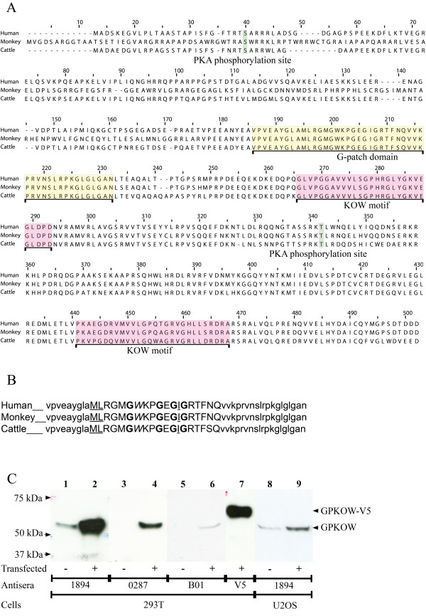
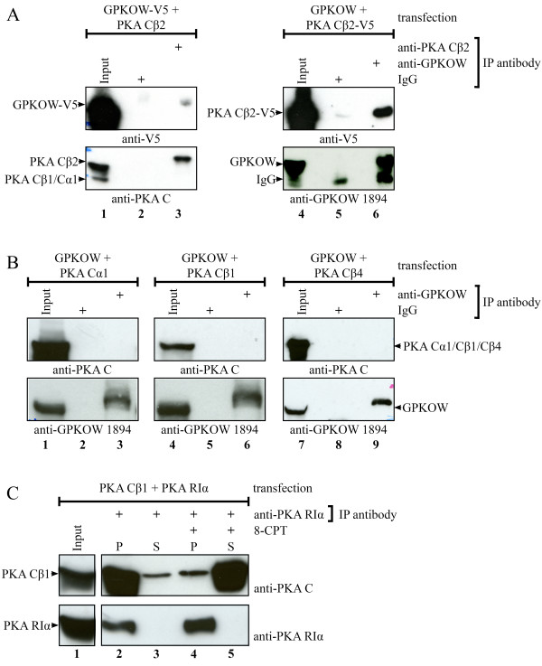
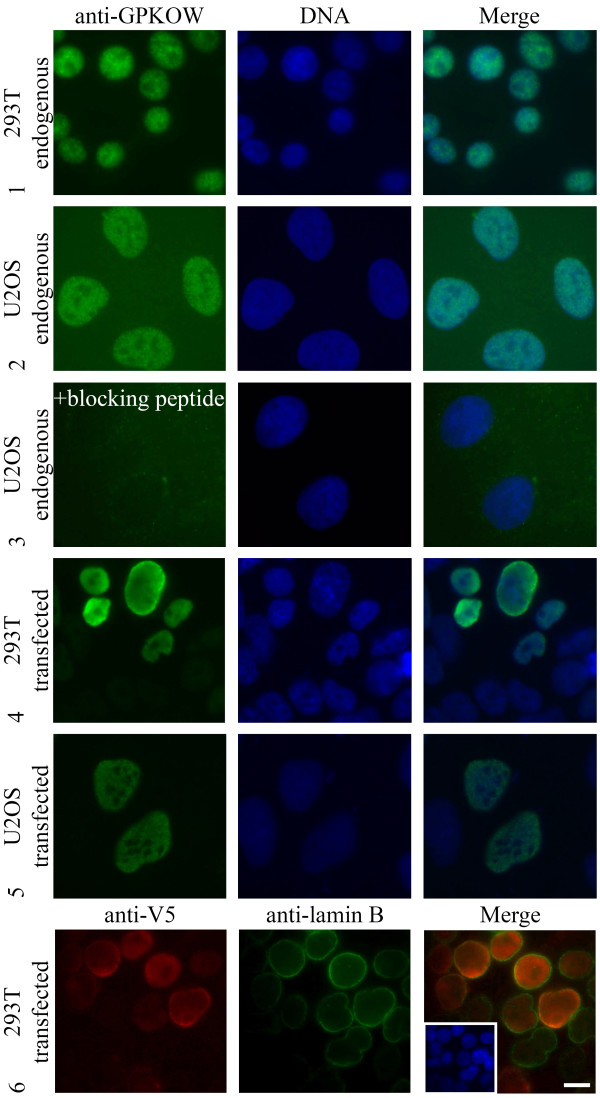
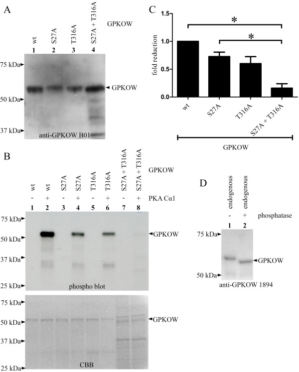
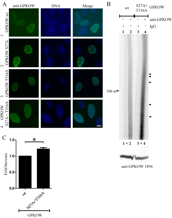
Results: Using yeast two-hybrid screening with the PKA C subunit Cbeta2 as bait, we identified the G-patch domain and KOW motifs-containing protein (GPKOW), also known as the T54 protein or MOS2 homolog, as an interaction partner for Cbeta2. We demonstrate that GPKOW, which contains one G-patch domain and two KOW motifs, is a nuclear RNA-binding protein conserved between species. GPKOW contains two sites that are phosphorylated by PKA in vitro. By RNA immunoprecipitation and site directed mutagenesis of the PKA phosphorylation sites we revealed that GPKOW binds RNA in vivo in a PKA sensitive fashion.

Conclusion: GPKOW is a RNA-binding protein that binds RNA in a PKA regulated fashion. Together with our previous results demonstrating that PKA regulates pre-mRNA splicing, our results suggest that PKA phosphorylation is involved in regulating RNA processing at several steps.


Figures

Figure 1
Figure 1
Sequence, domains and expression of GPKOW. A. Sequence alignment of GPKOW from human (NCBI NP_056513.2, upper line), monkey (NCBI XP_001105674, middle line) and cattle (NCBI XP_616889, lower line). The G-patch domain is indicated in yellow and the two KOW motifs are indicated in red. Two potential PKA phosphorylation sites, S27 and T316 (according to human GPKOW), are indicated in green. B. The G-patch domain of GPKOW as defined in the ELM database. Upper case letters illustrate the G-patch consensus sequence, where bold letters are glycines, underscored letters are hydrophobic residues and aromatic amino acids are marked in italic. C. 293T cells were left untreated (lanes 1, 3 and 5) or transfected with full-length GPKOW (lanes 2, 4 and 6) or GPKOW with a C-terminal V5-tag (lane 7). U2OS cells were left untreated (lane 8) or transfected with full-length GPKOW (lane 9). Twenty hours post transfection the cells were harvested and lysed. Eighteen μg (lanes 1-4 and 7), 36 μg (lane 5-6) or 46 μg (lanes 8-9) of protein was loaded per lane, separated by SDS-PAGE (10% gels) and analyzed by immunoblotting using three different GPKOW antibodies, 1894 (lanes 1-2 and 8-9), 0287 (lanes 3-4) and B01 (lanes 5-6) or V5 antibody (lane 7). One representative immunoblot from each experiment is presented.
Figure 2
Figure 2
Co-immunoprecipitations of GPKOW and PKA Cβ2. A. 293T cells were transfected with native PKA Cβ2 and GPKOW with a C-terminal V5-tag (lanes 1-3) or with native GPKOW and PKA Cβ2 with a C-terminal V5-tag (lanes 4-6). Twenty hours post transfection the cells were harvested and lysed. The lysates were adjusted to equal protein concentration for each experiment and precleared with magnetic beads before input samples were collected (lanes 1 and 4). The lysates were subjected to immunoprecipitations using anti-PKA Cβ2 (lane 3), anti-GPKOW (lane 6) or rabbit IgG (lanes 2 and 5) and magnetic beads. All samples were analyzed by SDS-PAGE and immunoblotting using anti-PKA C (lower panel lanes 1-3), anti-GPKOW (lower panel lanes 4-6) or anti-V5 (upper panel). One representative experiment clearly showing interaction is presented. B. 293T cells were co-transfected with native GPKOW and PKA Cα1 (lanes 1-3), PKA Cβ1 (lanes 4-6) or PKA Cβ4 (lanes 7-9) and treated as described in A. Input samples (lane1, 4 and 7) and immunoprecipitations with anti-GPKOW (lane 3, 6 and 9) or rabbit IgG (lanes 2, 5 and 8) were immunoblotted with anti-GPKOW (lower panel) or anti-PKA C (upper panel). One representative experiment is presented. C. 293T cells were co-transfected with PKA Cβ1 and PKA RIα, treated as described in A and immunoprecipitated with anti-PKA RIα (lanes 2-5). One pellet were treated with the cAMP analog, 8-CPT (lanes 4-5), and all pellets (P) and supernatants (S) were analyzed by SDS-PAGE and immunoblotting using anti-PKA C (upper panel) or anti-PKA RIα (lower panel). One representative experiment is shown.
Figure 3
Figure 3
Localization of GPKOW. Immunofluorescence analysis of paraformaldehyde fixated 293T (rows 1, 4 and 6) and U2OS cells (rows 2, 3 and 5) stained with anti-GPKOW 1894 (rows 1-5, green), anti-V5 (row 6, red) or anti-lamin B (row 6, green). In the fourth row the antibody was preincubated with antigen (+ blocking peptide) before incubation. The three lower rows show anti-GPKOW and anti-V5 staining of transfected full-length (rows 4-5) or V5-tagged (row 6) GPKOW. Nuclear DNA was visualized with Hoechst (rows 1-6, blue). Merged photos to the right. Scale bar, 10 μM.
Figure 4
Figure 4
GPKOW binds RNA in vivo. A. 293T cells were harvested and exposed to UV irradiation (254 nm/200 mJ). The cells were lysed and treated with T1 RNase (0.048 U/μl) for 8 min followed by immunoprecipitation with Dynabeads conjugated with anti-GPKOW1894 (+ anti-GPKOW, lane 2) or rabbit IgG (+ IgG, lane 1). Immunoprecipitated samples were dephosphorylated with calf intestinal phosphatase (0.038 U/μl). The RNA was labeled with γ32-P ATP by PNK (0.5 U/μl) phosphorylation. All samples were separated on a denaturating (7 M urea) 6% PAA gel and subjected to autoradiography. The arrows indicate specific RNA species. The lower panel shows immunoreactive GPKOW from the cell lysate used (one representative lane is shown). B. Unsaturated images from the Typhoon 9410 phosphoimager (GE Healthcare Life Sciences) were quantified using the histogram function in Adobe Photoshop. The lanes were manually defined with identical frames. The statistical analysis was performed with paired students T-test in GraphPad Prism. The asterisk indicates a significant difference between the columns with a p-value < 0.05 (n = 4).
Figure 5
Figure 5
PKA phosphorylates GPKOW at S27 and T316 in vitro. A. Recombinant GPKOW proteins were expressed in bacteria and purified on a HisTrap column followed by an ion exchange column. GPKOW wt (lane 1), GPKOW S27A (lane 2), GPKOW T316A (lane 3) and GPKOW S27A+T316A (lane 4) were all recognized by anti-GPKOW B01 after separation on SDS-PAGE and immunoblotting. One representative immunoblot is shown. B. Purified GPKOW wt (lanes 1 and 2), single- (lanes 3-6) or double-mutated GPKOW (lanes 7 and 8) were incubated with active (+) or heat inactivated (-) PKA Cα1 (7.4 ng) and γ-32P-ATP in a reaction buffer. The samples were analyzed by SDS-PAGE and autoradiography. The CBB staining in the lower panel shows the amount of the different proteins. C. Unsaturated images from Syngene G-box and Typhoon 9400 phosphoimager were quantified by Genetools (Syngene) and the statistical analysis was performed with paired students T-test in GraphPad Prism. The asterisk indicates a significant difference between the columns with a p-value < 0.01 (n = 3). D. 293T cells were harvested and lysed. The lysates were adjusted to equal protein concentration, precleared with magnetic beads before immunoprecipitation using anti-GPKOW (lane 1-2) and magnetic beads. Sample 2 was treated with alkaline phosphatase for 30 minutes before both samples were analyzed by SDS-PAGE and immunoblotting using anti-GPKOW. One representative experiment is shown (n = 3).
Figure 6
Figure 6
GPKOWs ability to bind RNA is sensitive to mutations of its PKA phosphorylation sites. A. U2OS cells were transfected with GPKOW wt (row 1), GPKOW S27A (row 2), GPKOW T316A (row 3) or GPKOW S27A+T316A (row 4). Cells were fixated with paraformaldehyde 20 hours later and subjected to immunofluorescence analysis using anti-GPKOW1894 (left, green). Nuclear DNA was visualized with Hoechst (middle, blue). Merged photos to the right. Scale bar 10 μM B. 293T cells transfected with GPKOW wt (lanes 1 and 2) or GPKOW S27A + T316A (lanes 3 and 4) were harvested and exposed to UV irradiation at 254 nm (200 mJ). The cells were lysed and immunoprecipitated with Dynabeads conjugated with either rabbit IgG (+ IgG, lanes 1 and 3) or anti-GPKOW1894 (+ anti-GPKOW, lanes 2 and 4). Immunoprecipitated samples were dephosphorylated with calf intestinal phosphatase (0.038 U/μl) followed by labeling of RNA with PNK (0.5 U/μl) phosphorylation. The samples were separated in a denaturating (7 M urea) 6% polyacrylamide gel and subjected to autoradiography. The arrows indicate specific RNA species. The lower panel shows immunoreactive GPKOW from the cell lysate used (one representative lane is shown). n = 4 C. Unsaturated images from the Typhoon 9410 phosphoimager (GE Healthcare Life Sciences) were quantified using the histogram function in Adobe Photoshop. The lanes were manually defined with identical frames. The statistical analysis was performed with paired students T-test in GraphPad Prism. The asterisk indicates a significant difference between the columns with a p-value < 0.01 (n = 4).

References

    1. Hui J. Regulation of mammalian pre-mRNA splicing. Sci China C Life Sci. 2009;52:253–260. doi: 10.1007/s11427-009-0037-0. - DOI - PubMed
    1. Glisovic T, Bachorik JL, Yong J, Dreyfuss G. RNA-binding proteins and post-transcriptional gene regulation. FEBS Lett. 2008;582:1977–1986. doi: 10.1016/j.febslet.2008.03.004. - DOI - PMC - PubMed
    1. Burd CG, Dreyfuss G. RNA binding specificity of hnRNP A1: significance of hnRNP A1 high-affinity binding sites in pre-mRNA splicing. EMBO J. 1994;13:1197–1204. - PMC - PubMed
    1. Keene JD. Minireview: global regulation and dynamics of ribonucleic Acid. Endocrinology. 2010;151:1391–1397. doi: 10.1210/en.2009-1250. - DOI - PMC - PubMed
    1. Keene JD. RNA regulons: coordination of post-transcriptional events. Nat Rev Genet. 2007;8:533–543. doi: 10.1038/nrg2111. - DOI - PubMed