. 2011 Aug 31:10:106.
doi: 10.1186/1476-4598-10-106.

Mesothelin confers pancreatic cancer cell resistance to TNF-α-induced apoptosis through Akt/PI3K/NF-κB activation and IL-6/Mcl-1 overexpression

Affiliations

Mesothelin confers pancreatic cancer cell resistance to TNF-α-induced apoptosis through Akt/PI3K/NF-κB activation and IL-6/Mcl-1 overexpression

Uddalak Bharadwaj et al. Mol Cancer. .

Abstract

Background: Previous studies showed that mesothelin (MSLN) plays important roles in survival of pancreatic cancer (PC) cells under anchorage dependent/independent conditions as well as resistance to chemotherapy. The recent success of intratumorally-injected adeno-encoded, chemo/radiation-inducible-promoter driven hTNF-α, (TNFerade) + gemcitabine in pre-clinical models of PC have renewed interest in use of TNF-α as a therapeutic component. To help find additional factors which might affect the therapy, we examined the resistance of MSLN-overexpressing pancreatic cancer cell lines to TNF-α-induced growth inhibition/apoptosis.

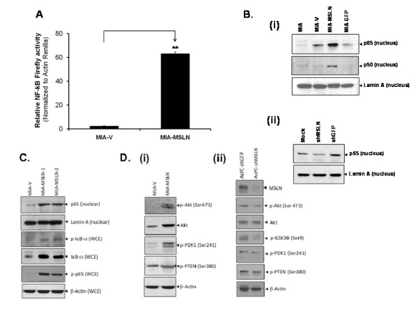
Methods: Stable MSLN overexpressing MIA PaCa-2 cells (MIA-MSLN), stable MSLN-silenced AsPC-1 cells (AsPC-shMSLN) and other pancreatic cells (MIA-PaCa2, Panc 28, Capan-1, BxPC3, PL 45, Hs 766T, AsPC-1, Capan-2, Panc 48) were used. NF-κB activation was examined by western blots and luciferase reporter assay. TNF-α induced growth inhibition/apoptosis was measured by MTT, TUNEL assay and caspase activation. IL-6 was measured using luminex based assay.

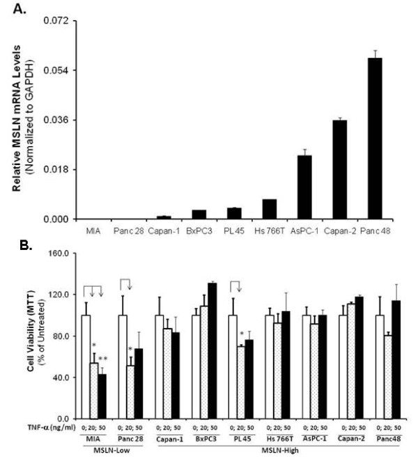
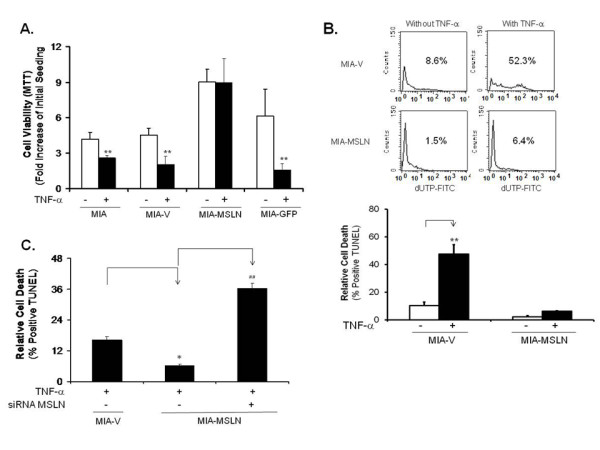
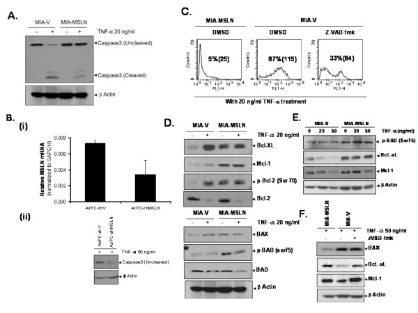
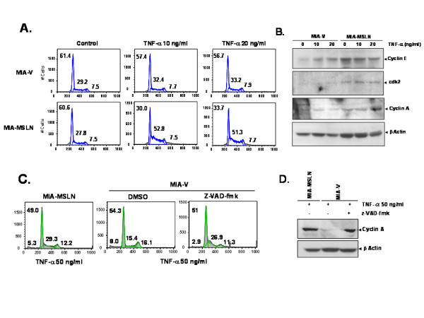
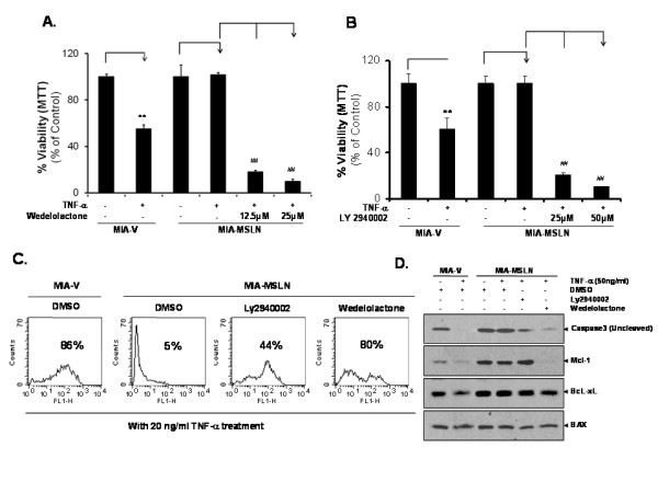
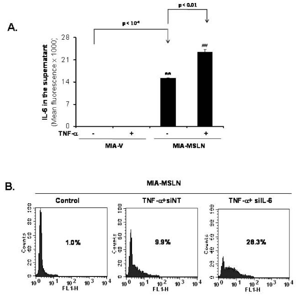
Results: Compared to low endogenous MSLN-expressing MIA PaCa-2 and Panc 28 cells, high endogenous MSLN-expressing Capan-1, BxPC3, PL 45, Hs 766T, AsPC-1, Capan-2, Panc 48 cells were resistant to TNF-α induced growth inhibition. Stable MSLN overexpressing MIA-PaCa2 cells (MIA-MSLN) were resistant to TNF-α-induced apoptosis while stable MSLN-silenced AsPC1 cells (AsPC-shMSLN) were sensitive. Interestingly, TNF-α-treated MIA-MSLN cells showed increased cell cycle progression and cyclin A induction, both of which were reversed by caspase inhibition. We further found that MIA-MSLN cells showed increased expression of anti-apoptotic Bcl-XL and Mcl-1; deactivated (p-Ser75) BAD, and activated (p-Ser70) Bcl-2. Constitutively activated NF-κB and Akt were evident in MIA-MSLN cells that could be suppressed by MSLN siRNA with a resultant increase in sensitivity of TNF-α induced apoptosis. Blocking NF-κB using IKK inhibitor wedelolactone also increased sensitivity to TNF-α-mediated cytotoxicity with concomitant decrease in Mcl-1. Blocking Akt using PI3K inhibitor also had a likewise effect presumably affecting cell cycle. MIA-MSLN cells produced increased IL-6 and were increased furthermore by TNF-α treatment. SiRNA-silencing of IL-6 increased TNF-α sensitivity of MIA-MSLN cells.

Conclusions: Our study delineates a MSLN-Akt-NF-κB-IL-6-Mcl-1 survival axis that may be operative in PC cells, and might help cancer cells' survival in the highly inflammatory milieu evident in PC. Further, for the success of TNFerade + gemcitabine to be successful, we feel the simultaneous inhibition of components of this axis is also essential.


Figures

Figure 1
Figure 1
MSLN expression level positively correlates with protection of pancreatic cancer cell lines from TNF-α induced apoptosis. (A). Evaluation of MSLN mRNA expression in PC cell lines. Relative MSLN mRNA levels in pancreatic cancer cell lines MIA PaCa-2, Panc28, Capan-1, BxPC3, PL 45, Hs 766T, AsPC-1, Capan-2 and Panc 48 cells. Total mRNA from the cell lines were reverse transcribed and tested for MSLN expression by Real Time PCR. The results depicted denote MSLN mRNA levels in each cell line normalized to the GAPDH mRNA level. Relative mRNA level is presented as 2^[Ct(GAPDH)-Ct(MSLN)]. The bars denote SD of duplicate data. (B). A panel of PC cells' viability with or without TNF-α treatment. Cells were seeded in 96-well plates, serum-starved for 24 h, and then cultured in serum free medium ± 20/50 ng/ml of TNF-α for 72 h. Viable cells were quantitated by using MTT assay. Relative fold increase in viability is plotted along Y axis. Data plotted show means of triplicate wells.
Figure 2
Figure 2
MSLN overexpression protects PC cells from TNF-α induced cell viability decrease and apoptosis. (A). MIA-MSLN and control cells viability assay with or without TNF-α treatment. About 3000 cells were plated in 96 well plates, serum starved for 24 h and treated with TNF-α at 20 ng/ml for 96 h after which viability was measured by MTT. Relative increase in viability was measured by dividing viability at a time point by viability of same cells at day 0 (day after plating) and is plotted along Y axis. Data plotted show mean of triplicate wells. Bars denote s.d. of triplicate data. *, # denote p < 0.05, **, ## denote p < 0.01, compared with controls by using student t test. (B). MIA-MSLN cells are resistant to TNF-α induced apoptosis using TUNEL assay. Cells were treated ± 20 ng/ml of TNF-α for 72 h and apoptosis was measured by TUNEL assay. Representative flow histograms show percentage of dUTP-FITC-positive apoptotic cells. Lower panel shows mean number of TUNEL positive cells (duplicate wells). (C). siRNA-silencing MSLN renders MIA-MSLN cells become TNF-α-sensitive measured by TUNEL assay. MIA-V/MIA-MSLN ells were plated in 6 well plates, transfected with siRNA, 24 h after transfection media was replaced by growth media, cells were serum starved and treated with TNF-α at 20 ng/ml for 72 h after which cells were collected, fixed and tested for DNA strand breaks by TUNEL assay. Bars denote s.d. of duplicate data. *, # denote p < 0.05, **, ## denote p < 0.01, compared with controls by using student t test.
Figure 3
Figure 3
MSLN overexpression makes PC cells resistant to TNF-α induced apoptosis through reduced Caspase 3 cleavage and up regulated anti-apoptotic proteins. (A). Reduced Caspase 3 cleavage in MIA-MSLN cells by 20 ng/ml of TNF-α treatment for 72 h. (B). Silencing endogenous MSLN in AsPC-3 cells renders it sensitive to TNF-α induced apoptosis through increased Caspase 3 cleavage. Panel (i) shows the reduction of MSLN expression level in AsPC-shMSLN stable cell line. Panel (ii) shows the increased caspase 3 cleavage upon TNF-α treatment. (C). Pre-treatment with pan-caspase inhibitor zVAD-fmk renders MIA-V cells resistant to TNF-α-induced apoptosis. Cells were treated with 20 ng/ml of TNF-α for 72 h and then stained with TUNEL assay. (D). Pro and anti-apoptotic molecules expression in MIA-V/MIA-MSLN cells with or without treatment of 20 ng/ml TNF-α assayed by Western blot. (E). Effect of different TNF-α doses on selected pro/anti-apoptotic molecules expression in MIA-V/MIA-MSLN cells. (F). Effect of zVAD-fmk pre-treatment on various pro and anti-apoptotic molecules expression in MIA-V/MIA-MSLN cells after treatment with 50 ng/ml of TNF-α for 72 h.
Figure 4
Figure 4
TNF-α induces S phase progression and induction of cyclin A in MIA-MSLN cells. (A). Cell cycle analysis of MIA-V/MIA-MSLN cells treated with 10/20 ng/ml of TNF-α for 24 h. (B). Cell cycle related molecules detection by western blot using the lysates from cells treated as in (A). (C). Cell cycle analysis of MIA-V/MIA-MSLN cells after pre-treatment with zVAD-fmk and then treated with 50 ng/ml of TNF-α for 72 h. (D). Cyclin A detection by western blot using the cell lysates from cells treated as in (C).
Figure 5
Figure 5
MSLN overexpression is associated with activation of NF-κB and Akt activity in PC cells. (A). Enhanced NF-κB reporter gene activity in MIA-MSLN cells. Data plotted was measured as ratio of co-transfected firefly-NF-κB reporter normalized to Actin-Renilla. ** denote p < 0.01, compared with controls, t test. (B). Nuclear protein from MIA-MSLN and control cells were subjected to western blot to detect nuclear-translocated p65, p50 and nuclear envelop protein Lamin A (panel i). Panel ii showed inhibited nuclear protein translocation when MIA-MSLN cells were transfected with MSLN specific shRNA encoding plasmid or control plasmids. (C). Whole cell extract (WCE) or nuclear protein from MIA-V and two pools of MIA-MSLN cells were probed for various IκB-α degradation pathway proteins in western blot. (D). Whole protein from MIA-V/MIA-MSLN (panel i) and from the AsPC-1 shMSLN and control cells (panel ii) were probed for various PI3k-Akt pathway proteins. Note: (panel i) data has individual lanes combined from a single electrophoresis gel.
Figure 6
Figure 6
Inhibiting NF-κB/Akt pathway or endogenous IL-6 abrogates MIA-MSLN cells resistance to TNF-α-induced apoptosis. (A). MIA-MSLN cell viability was reduced upon pre-treatment with IKK inhibitor wedelolactone and then 20 ng/ml of TNF-α for 72 h. (B). MIA-MSLN cell viability was reduced upon pre-treatment with PI3K inhibitor Ly 2940002 and then 20 ng/ml of TNF-α for 72 h. (C). Cell apoptosis was determined by TUNEL assay in samples treated as in (A) and (B). (D). Proteins from similarly treated cells were subjected to western blot to detect caspase 3 cleavage and key pro/anti-apoptotic molecules.
Figure 7
Figure 7
High levels of IL-6 in MIA-MSLN cells plays important role in resistance to TNF-α-mediated cytotoxicity. (A). Elevated constitutive and TNF-α induced IL-6 expression in MIA-MSLN cells using Luminex-based IL-6 assay kit. Y-axis represents fluorescence corresponding to IL-6-levels. Bars denote SD of duplicate data. (B). Inhibiting endogenous IL-6 by siRNA sensitizes MIA-MSLN cells to TNF-α-induced apoptosis. MIA-MSLN cells were either mock-transfected (control) or transfected with non-targeting (siNT) or IL-6 specific siRNA pools (siIL-6) and treated with 20 ng/ml of TNF-α for 72 h and analyzed for TUNEL positivity. Flow histograms show percentage of dUTP-FITC-positive apoptotic cells. Plots are representatives of three independent experiments.

Similar articles

Cited by

References

    1. Bharadwaj U, Li M, Chen C, Yao Q. Mesothelin-induced pancreatic cancer cell proliferation involves alteration of cyclin E via activation of signal transducer and activator of transcription protein 3. Mol Cancer Res. 2008;6:1755–1765. doi: 10.1158/1541-7786.MCR-08-0095. - DOI - PMC - PubMed
    1. Hassan R, Ho M. Mesothelin targeted cancer immunotherapy. Eur J Cancer. 2008;44:46–53. doi: 10.1016/j.ejca.2007.08.028. - DOI - PMC - PubMed
    1. Li M, Bharadwaj U, Zhang R, Zhang S, Mu H, Fisher WE, Brunicardi FC, Chen C, Yao Q. Mesothelin is a malignant factor and therapeutic vaccine target for pancreatic cancer. Mol Cancer Ther. 2008;7:286–296. doi: 10.1158/1535-7163.MCT-07-0483. - DOI - PMC - PubMed
    1. Uehara N, Matsuoka Y, Tsubura A. Mesothelin promotes anchorage-independent growth and prevents anoikis via extracellular signal-regulated kinase signaling pathway in human breast cancer cells. Mol Cancer Res. 2008;6:186–193. doi: 10.1158/1541-7786.MCR-07-0254. - DOI - PubMed
    1. Cheng WF, Huang CY, Chang MC, Hu YH, Chiang YC, Chen YL, Hsieh CY, Chen CA. High mesothelin correlates with chemoresistance and poor survival in epithelial ovarian carcinoma. Br J Cancer. 2009;100:1144–1153. doi: 10.1038/sj.bjc.6604964. - DOI - PMC - PubMed

Publication types

MeSH terms

Substances