. 2011 Aug 31:11:52.
doi: 10.1186/1471-213X-11-52.

Arx and Nkx2.2 compound deficiency redirects pancreatic alpha- and beta-cell differentiation to a somatostatin/ghrelin co-expressing cell lineage

Affiliations

Arx and Nkx2.2 compound deficiency redirects pancreatic alpha- and beta-cell differentiation to a somatostatin/ghrelin co-expressing cell lineage

Simon Kordowich et al. BMC Dev Biol. .

Abstract

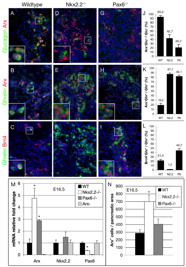
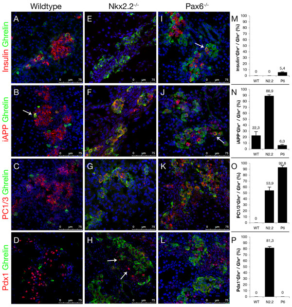
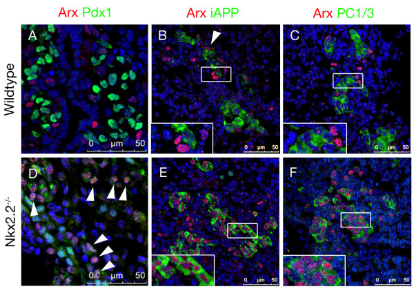
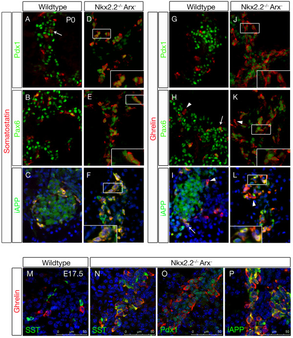
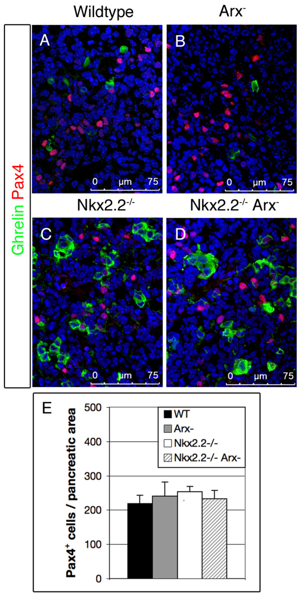
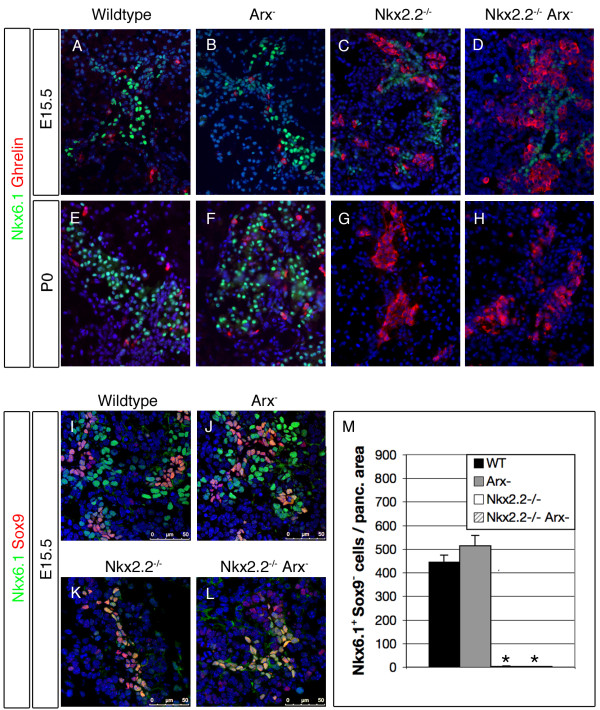
Background: Nkx2.2 and Arx represent key transcription factors implicated in the specification of islet cell subtypes during pancreas development. Mice deficient for Arx do not develop any alpha-cells whereas beta- and delta-cells are found in considerably higher numbers. In Nkx2.2 mutant animals, alpha- and beta-cell development is severely impaired whereas a ghrelin-expressing cell population is found augmented.Notably, Arx transcription is clearly enhanced in Nkx2.2-deficient pancreata. Hence in order to precise the functional link between both factors we performed a comparative analysis of Nkx2.2/Arx single- and double-mutants but also of Pax6-deficient animals.

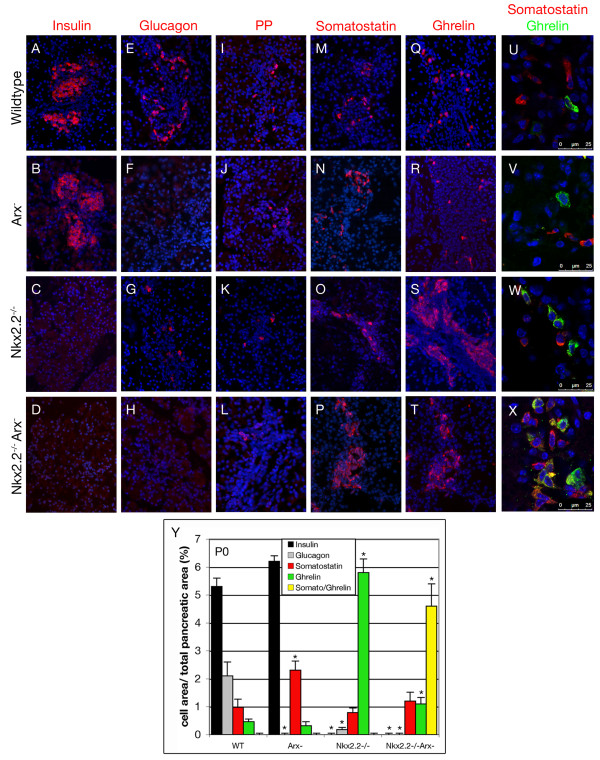
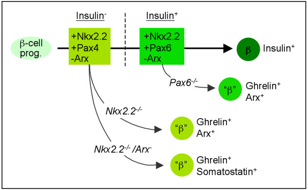
Results: We show that most of the ghrelin+ cells emerging in pancreata of Nkx2.2- and Pax6-deficient mice, express the alpha-cell specifier Arx, but also additional beta-cell related genes. In Nkx2.2-deficient mice, Arx directly co-localizes with iAPP, PC1/3 and Pdx1 suggesting an Nkx2.2-dependent control of Arx in committed beta-cells. The combined loss of Nkx2.2 and Arx likewise results in the formation of a hyperplastic ghrelin+ cell population at the expense of mature alpha- and beta-cells. Surprisingly, such Nkx2.2-/-Arx- ghrelin+ cells also express the somatostatin hormone.

Conclusions: Our data indicate that Nkx2.2 acts by reinforcing the transcriptional networks initiated by Pax4 and Arx in early committed beta- and alpha-cell, respectively. Our analysis also suggests that one of the coupled functions of Nkx2.2 and Pax4 is to counteract Arx gene activity in early committed beta-cells.


Figures

Figure 1
Figure 1
Expression of Arx and Brn4 in E16.5 pancreata of wild type, Nkx2.2-/- and Pax6-/- mice. (A, B) Arx is expressed in wild type glucagon+ cells. (A, inset) Moreover, Arx+/glucagon- cells are detectable at this stage. (B, C, inset) Arx or Brn4 can be detected in a subset of ghrelin+ cells. (D, inset) In Nkx2.2-mutant pancreata some of the remaining glucagon+ cells do not express Arx. (E, inset, F) Most Nkx2.2-deficient ghrelin+ cells express Arx but only rarely Brn4. (G, H) Arx is expressed in pancreata of Pax6-/- mice and co-localizes with most ghrelin+ cells. Scattered Arx+/ghrelin- and ghrelin+/Arx- cells can be detected. (I, inset) Brn4 is expressed in a subset of ghrelin+ cells and labels a ghrelin- cell population in Pax6-deficient mice. Counterstaining with DAPI. (J-L) The percentages of Arx+/glucagon+, Arx+/ghrelin+ and Brn4+/ghrelin+ cells in pancreata of wild type (WT), Nkx2.2-/- (N2.2) and Pax6-/- (P6) mice within the total glucagon+ and ghrelin+ cell populations, respectively, are depicted. (M) Quantitative RT-PCR of Arx, Pax6 and Nkx2.2 mRNA in wild type, Nkx2.2-/-, Pax6-/- and Arx- E16.5 pancreas. (N) Quantification of Arx+ cells at E16.5 in pancreata of wild type, Nkx2.2-/- and Pax6-/- mice (n = 3 pancreas per genotype). Single-factor ANOVA coupled to Newman-Keuls test was applied and p-values < 0,05 were assessed as statistically significant compared to wild type. Statistical significance is achieved with the assumption of a normal distribution. Error bars represent SEM. (original magnification: 400×, Insets magnification: 4×).
Figure 2
Figure 2
Co-detection of ghrelin and insulin, iAPP, PC1/3 or Pdx1 in pancreata of wild type, Nkx2.2-/- and Pax6-/- mice at E17.5. (A-D) Wild type ghrelin+ cells do not express insulin, PC1/3 or Pdx1. However, scattered ghrelin+ cells express iAPP (Arrow). (E-H) No insulin reactive cells are detectable in Nkx2.2-deficient pancreata. The majority of ghrelin+ cells express iAPP and low levels of Pdx1. Scattered iAPP+/ghrelin- (F) and Pdx1+/ghrelin- cells (H, Arrows) are detectable. (G) PC1/3 is associated with a subpopulation of ghrelin-expressing cells in Nkx2.2-deficient pancreata. (I-L) In pancreata deficient for Pax6 the population of ghrelin+ cells does not express insulin or iAPP except in very rare instances (Arrows in I+J). Almost all Pax6-deficient ghrelin+ cells express PC1/3 but none produce Pdx1. Counterstaining with DAPI. (M-P) The percentages of insulin+/ghrelin+, iAPP+/ghrelin+, PC1/3+/ghrelin+ and Pdx1+/ghrelin+ cells in wild type (WT), Nkx2.2-/- (N2.2) and Pax6-/- (P6) mice within the total ghrelin+ cell population are depicted. Pictures were taken using confocal microscopy.
Figure 3
Figure 3
Co-detection of Arx and Pdx1, iAPP or PC1/3 in pancreata of wild type and Nkx2.2-/- mice at E17.5. (A-C) Wild type Arx+ cells do not express Pdx1 (A) or PC1/3 (C, inset). Most Arx+ cells do not express iAPP (B), except in rare instances (Arrowhead). (D-F) The majority of Arx+ cells in Nkx2.2-/- pancreata express Pdx1low (Arrows), iAPP (E, inset) and PC1/3 (F, inset). Counterstaining with DAPI. Pictures were taken using confocal microscopy (Insets magnification: 4×).
Figure 4
Figure 4
Expression of endocrine hormones in pancreata of wild type, Arx-, Nkx2.2-/- and Nkx2.2-/-Arx- newborns. Insulin (A-D), glucagon (E-H), pancreatic polypeptide (I-L), somatostatin (M-P) and ghrelin (Q-T) immunofluorescence-detection at P0. (D, H) Note that both beta- and alpha-cells lack in Nkx2.2-/-Arx- double-mutant mice (original magnification: 200×). (U-X) Co-detection of ghrelin and somatostatin in P0 pancreata of wild type, Arx-, Nkx2.2-/- and Nkx2.2-/-Arx- mice. Somatostatin+ cells and ghrelin+ cells label distinct cell populations in wild type (U), Arx- (V) and Nkx2.2-deficient (W) pancreata. (X) However, in Nkx2.2-/-Arx- double mutant mice a population of ghrelin+/somatostatin+ co-positive cells is detectable besides single-hormone-expressing ghrelin+ or somatostatin+ cells. Pictures in U-X were taken using confocal microscopy. Counterstaining with DAPI. (Y) Quantification of hormone producing cells (without PP-cells) in pancreata of wild type, Arx-, Nkx2.2-/- and Nkx2.2-/-Arx- mice at P0. Bars represent percentage of fluorescent cell area per total pancreatic area. n = 3 pancreas per genotype. Single-factor ANOVA coupled to Newman-Keuls test was applied and p-values < 0,05 were assessed as statistically significant compared to wild type. Statistical significance is achieved with the assumption of a normal distribution. Error bars represent SEM.
Figure 5
Figure 5
Expression of Pdx1, Pax6 and iAPP in pancreata of wild type and Nkx2.2-/-Arx- newborns. (A) In wild type pancreata, a subpopulation of delta-cells expresses low levels of Pdx1 (Arrow) while the bulk does not. (B, C) Wild type delta-cells express Pax6 and iAPP. (D, inset) Notably, in Nkx2.2-/-Arx- mice almost all somatostatin+ cells display low levels of Pdx1. (E, F, insets) Nkx2.2-/-Arx- somatostatin+ cells are immunoreactive for iAPP and low levels of Pax6. (G) Wild type epsilon-cells do not express Pdx1. (H, I) Pax6 or iAPP can be detected in a subset of wild type ghrelin+ cells (Arrows); a few ghrelin+ cells are negative for Pax6 or iAPP (Arrowheads). (J, K) In Nkx2.2-/-Arx- pancreata, the majority of ghrelin+ cells are found positive for low levels of Pdx1 and Pax6; scattered ghrelin+ cells do not express Pax6 (K, Arrowhead). (L, inset) Islet amyloid polypeptide is detectable in the majority of ghrelin+ cells; some are negative for iAPP (Arrowhead). Pictures in D, E, J and K have been captured using a 2-fold exposure time compared to respective control images in order to allow for a clear detection of Pdx1 and Pax6 (original magnification 800×). C, F, I and L are counterstained with DAPI. (M-N) Confocal images of E17.5 wild type and Nkx2.2/Arx double-mutant pancreata. Similar to the expression patterns in newborns, Nkx2.2/Arx double-deficient ghrelin+ cells express somatostatin, Pdx1low and iAPP at E17.5.
Figure 6
Figure 6
Pax4 expression in pancreata of wild type, Arx-, Nkx2.2-/- and Nkx2.2-/-/Arx- at E16.5. (A-D) Pax4-expressing cells are present in normal numbers at E16.5 in pancreata of wild type, Arx-, Nkx2.2-/- and Nkx2.2-/-/Arx- mice. Notably, no co-expression of Pax4 and ghrelin is detectable in wild type or mutant pancreata. Pictures were taken using confocal microscopy. (E) Quantification of Pax4+ cells at E16.5 in pancreata of wild type, Arx-, Nkx2.2-/- and Nkx2.2-/-Arx- mice (n = 3 pancreas per genotype). Single-factor ANOVA coupled to Newman-Keuls test was applied and p-values < 0,05 were assessed as statistically significant compared to wild type. Statistical significance is achieved with the assumption of a normal distribution. Error bars represent SEM.
Figure 7
Figure 7
Nkx6.1 expression is differentially dependent on Nkx2.2. (A-D) Nkx6.1 is broadly expressed in the pancreatic epithelium of wild type, Arx-, Nkx2.2-/- and Nkx2.2-/-Arx- mice at embryonic day 15.5. (E, F) In wild type and Arx-mutants, Nkx6.1 expression is maintained and detectable at P0. (G, H) However, in the absence of Nkx2.2 or Nkx2.2/Arx, a steady decrease in Nkx6.1+ cell numbers is detected from E15.5 on so that Nkx6.1+ cells are not detectable in pancreata of newborns. No co-expression of Nkx6.1 and ghrelin is detectable in wild type or mutant tissues at the investigated developmental time-points (original magnification 400×). (I-L) Nkx6.1+Sox9+ ductal/endocrine progenitors are present in all four genotypes at E15.5. In wild type and Arx-mutants Nkx6.1+Sox9- cells are detected in comparable numbers. (K, L) However, in the absence of Nkx2.2 or Nkx2.2 and Arx no such cells are detectable. Pictures in I-L were taken using confocal microscopy. (M) Quantification of Nkx6.1+Sox9- cell numbers in E15.5 pancreata of wild type, Arx-, Nkx2.2-/- and Nkx2.2-/-Arx- mice (n = 3 pancreas per genotype). Single-factor ANOVA coupled to Newman-Keuls test was applied and p-values < 0,05 were assessed as statistically significant compared to wild type. Statistical significance is achieved with the assumption of a normal distribution. Error bars represent SEM.
Figure 8
Figure 8
Schematic representation of transcription factor combinations guiding beta-cell differentiation. Following the loss of Nkx2.2 or Pax6, endocrine cells retain some beta-cell characteristics, such as the expression of iAPP, Pdx1 and/or PC1/3 (indicated as "β"). Rectangles describe transcription factor combinations required to maintain the embryonic beta-cell differentiation program pre- and post-insulin expression (light and dark green, respectively). Shortly after the activation of Ngn3, both Nkx2.2 and Pax4 are necessary for the expression of additional beta-cell specific TF's such as Nkx6.1 (not depicted). Concomitantly, this combination of TF's prevents the misexpression of Arx and ghrelin in committed beta-cells. In the absence of both Nkx2.2 and Arx, endocrine cells retain the same beta-cell features as noted in the absence of Nkx2.2 only, but then express ghrelin and somatostatin. Pax6 appears necessary for maintaining normal beta-cell identity at later stages of differentiation, probably shortly after insulin activation. At that stage Pax6 presumably takes over some of the functions previously exerted by Pax4, whose expression decreases.

References

    1. Adrian TE, Bloom SR, Hermansen K, Iversen J. Pancreatic polypeptide, glucagon and insulin secretion from the isolated perfused canine pancreas. Diabetologia. 1978;14:413–7. doi: 10.1007/BF01228136. - DOI - PubMed
    1. Heller RS, Jenny M, Collombat P, Mansouri A, Tomasetto C, Madsen OD, Mellitzer G, Gradwohl G, Serup P. Genetic determinants of pancreatic epsilon-cell development. Dev Biol. 2005;286:217–24. doi: 10.1016/j.ydbio.2005.06.041. - DOI - PubMed
    1. Korbonits M, Goldstone AP, Gueorguiev M, Grossman AB. Ghrelin--a hormone with multiple functions. Front Neuroendocrinol. 2004;25:27–68. doi: 10.1016/j.yfrne.2004.03.002. - DOI - PubMed
    1. Roncoroni L, Violi V, Montanari M, Muri M. Effect of somatostatin on exocrine pancreas evaluated on a total external pancreatic fistula of neoplastic origin. Am J Gastroenterol. 1983;78:425–8. - PubMed
    1. Wierup N, Svensson H, Mulder H, Sundler F. The ghrelin cell: a novel developmentally regulated islet cell in the human pancreas. Regul Pept. 2002;107:63–9. doi: 10.1016/S0167-0115(02)00067-8. - DOI - PubMed

Publication types

MeSH terms

Substances