. 2011 Sep 1:10:107.
doi: 10.1186/1476-4598-10-107.

Plumbagin inhibits invasion and migration of breast and gastric cancer cells by downregulating the expression of chemokine receptor CXCR4

Affiliations

Plumbagin inhibits invasion and migration of breast and gastric cancer cells by downregulating the expression of chemokine receptor CXCR4

Kanjoormana Aryan Manu et al. Mol Cancer. .

Abstract

Background: Increasing evidence indicates that the interaction between the CXC chemokine receptor-4 (CXCR4) and its ligand CXCL12 is critical in the process of metastasis that accounts for more than 90% of cancer-related deaths. Thus, novel agents that can downregulate the CXCR4/CXCL12 axis have therapeutic potential in inhibiting cancer metastasis.

Methods: In this report, we investigated the potential of an agent, plumbagin (5-hydroxy-2-methyl-1, 4-naphthoquinone), for its ability to modulate CXCR4 expression and function in various tumor cells using Western blot analysis, DNA binding assay, transient transfection, real time PCR analysis, chromatin immunoprecipitation, and cellular migration and invasion assays.

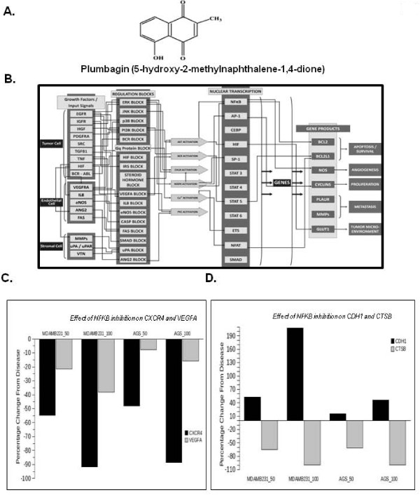
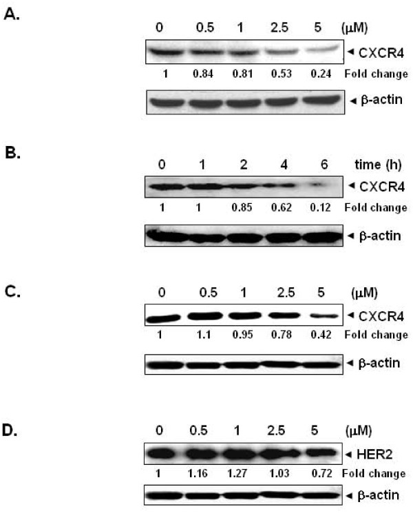
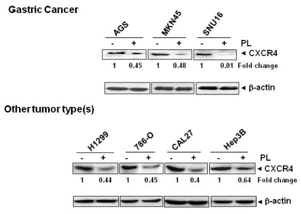
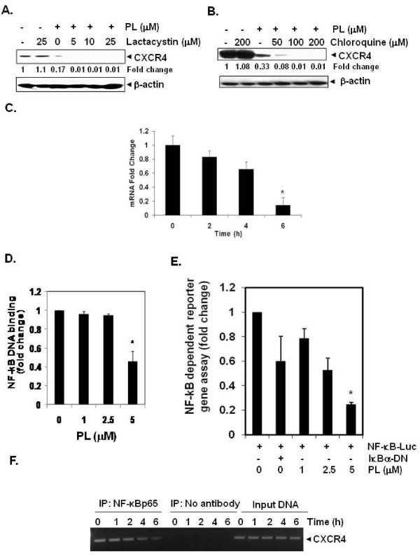
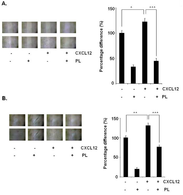
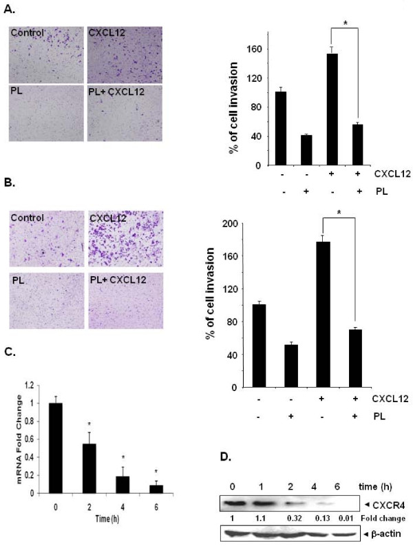
Results: We found that plumbagin downregulated the expression of CXCR4 in breast cancer cells irrespective of their HER2 status. The decrease in CXCR4 expression induced by plumbagin was not cell type-specific as the inhibition also occurred in gastric, lung, renal, oral, and hepatocellular tumor cell lines. Neither proteasome inhibition nor lysosomal stabilization had any effect on plumbagin-induced decrease in CXCR4 expression. Detailed study of the underlying molecular mechanism(s) revealed that the regulation of the downregulation of CXCR4 was at the transcriptional level, as indicated by downregulation of mRNA expression, inhibition of NF-κB activation, and suppression of chromatin immunoprecipitation activity. In addition, using a virtual, predictive, functional proteomics-based tumor pathway platform, we tested the hypothesis that NF-κB inhibition by plumbagin causes the decrease in CXCR4 and other metastatic genes. Suppression of CXCR4 expression by plumbagin was found to correlate with the inhibition of CXCL12-induced migration and invasion of both breast and gastric cancer cells.

Conclusions: Overall, our results indicate, for the first time, that plumbagin is a novel blocker of CXCR4 expression and thus has the potential to suppress metastasis of cancer.


Figures

Figure 1
Figure 1
Predictive In Silico Virtual tumor platform generated results. A, The chemical structure of plumbagin. B, The figure illustrates a high-level view of the maze of interactions and cross-talks present in the Virtual Tumor Cell platform. C, The figure illustrates the percentage reduction in CXCR4 and VEGFA with 50% and 100% inhibition of NF-κB in MDA-MB-231 (breast cancer) and AGS (gastric cancer) aligned virtual tumor cells. D, The figure illustrates the percentage reduction in metastatic tumor markers- CDH1 and Cathepsin B with 50% and 100% inhibition of NF-κB activation in the two tumor baselines.
Figure 2
Figure 2
Plumbagin suppresses CXCR4 expression in breast cancer cells. A, Plumbagin suppresses CXCR4 levels in a dose-dependent manner. MDA-MB-231 cells (1 × 10⁶) were treated with the indicated concentrations of plumbagin for 6 h. Whole-cell extracts were then prepared, and 30 μg of protein was resolved on SDS-PAGE, electrotransferred onto nitrocellulose membranes, and probed for CXCR4. The same blots were stripped and reprobed with β-actin antibody to show equal protein loading. B, Plumbagin suppresses CXCR4 levels in a time-dependent manner. MDA-MB-231 cells (1 × 10⁶) were treated with 5 μM plumbagin for the indicated times, after which Western blotting was done as described above. The same blots were stripped and reprobed with β-actin antibody to show equal protein loading. C, Plumbagin suppresses CXCR4 levels in HER2 overexpressing BT474 cells. BT474 cells (1 × 10⁶) were treated with the indicated concentrations of plumbagin for 6 h. Whole-cell extracts were then prepared, and 30 μg of protein was resolved on SDS-PAGE, electro-transferred onto nitrocellulose membranes, and probed for CXCR4. The same blots were stripped and reprobed with β-actin antibody to show equal protein loading. D, Effect of plumbagin on HER2 expression in BT474 cells. BT474 cells (1 × 10⁶) were treated with the indicated concentrations of plumbagin for 6 h, after which Western blotting for HER2 was done as described above. The same blots were stripped and reprobed with β-actin antibody to show equal protein loading. The representative results of three independent experiments are shown.
Figure 3
Figure 3
Plumbagin downregulates CXCR4 in different tumor cell types. Gastric cancer (AGS, MKN45, and SNU16), lung adenocarcinoma (H1299), renal cell carcinoma (786-O), oral adenosquamous carcinoma (CAL27), hepatocellular carcinoma (Hep3B) cells were incubated with 5 μM plumbagin for 6 h. Whole-cell extracts were prepared and analyzed by Western blot analysis using antibody against CXCR4. The same blots were stripped and reprobed with β-actin antibody to show equal protein loading. Representative results of three independent experiments are shown.
Figure 4
Figure 4
Plumbagin suppresses CXCR4 through modulation of its mRNA level. A and B, Plumbagin suppresses CXCR4, through lysosomal but not proteosomal degradation. Cells were treated with indicated concentrations of lactacystin or chloroquine for 1 h at 37°C, followed by treatment of 5 μM plumbagin for 6 h. Whole-cell extracts were prepared and analyzed by Western blot analysis using antibodies against CXCR4. The same blots were stripped and reprobed with β-actin antibody to show equal protein loading. Representative results of three independent experiments are shown. C, Plumbagin suppresses expression of CXCR4 mRNA in MDA-MB-231 cells. Cells were treated with 5 μM plumbagin for indicated times. Total RNA was isolated and analyzed by RT-PCR assay as described in Materials and Methods. 18S was shown to equal loading of total RNA. Representative results of three independent experiments are shown. D, Plumbagin inhibits NF-κB activation in MDA-MB-231 breast cancer cells. MDA-MB-231 cells were incubated with indicated concentrations of plumbagin for 2 h. The nuclear extracts were assayed for NF-κB activation by TransAM p65 transcription factor assay kit. E, MDA-MB-231 cells were transiently transfected with an NF-κB-luciferase plasmid and then treated with the indicated concentrations of plumbagin for 2 h. Cell supernatants were thereafter collected and assayed for luciferase activity as described in Materials and Methods. Representative results of three independent experiments are shown. Results are expressed as fold activity over the activity of the vector control. Bars indicate standard deviation. * indicates p value < 0.05). F, Plumbagin inhibits binding of NF-κB to the CXCR4 promoter. MDA-MB-231 cells were treated with 5 μM plumbagin for indicated time intervals and the proteins were cross-linked with DNA by formaldehyde and then subjected to ChIP assay using an anti-p65 antibody with the CXCR4 primer. Reaction products were resolved by electrophoresis.
Figure 5
Figure 5
Plumbagin suppresses migration of breast and gastric cancer cells. A, The wound-healing assay for evaluating the inhibitory effect of plumbagin on MDA-MB-231 cell migration. Confluent monolayers of MDA-MB-231 cells were scarred, and repair was monitored microscopically after 6 h of pre-treatment with 5 μM plumbagin before being exposed to 100 ng/mL CXCL12 for 24 h. Width of wound was measured at time zero and 24 h of incubation with and without plumbagin in the absence or presence of CXCL12 in RPMI medium containing 1% serum. The representative photographs showed the same area at time zero and after 48 h of incubation. Graphs, mean (n = 3); bars, SE. *, P < 0.05. B, The wound-healing assay for evaluating the inhibitory effect of plumbagin on AGS cell migration. Confluent monolayers of AGS cells were scarred, and repair was monitored microscopically after 6 h of pre-treatment with 5 μM plumbagin before being exposed to 100 ng/mL CXCL12 for 24 h. Width of wound was measured at time zero and 24 h of incubation with and without plumbagin in the absence or presence of CXCL12 in RPMI medium containing 1% serum. The representative photographs of three independent experiments showed the same area at time zero and after 48 h of incubation. Graphs, mean (n = 3); bars, SE. *, P < 0.05.
Figure 6
Figure 6
Plumbagin suppresses invasion in breast and gastric cancer cells. A, MDA-MB-231 (2 × 10⁵ cells) were seeded in the top-chamber of the Matrigel. After pre-incubation with or without plumbagin (5 μM) for 6 h, transwell chambers were then placed into the wells of a 24-well plate, in which we had added either the basal medium only or basal medium containing 100 ng/mL CXCL12 for 24 h. After incubation, they were assessed for cell invasion as described in Materials and Methods. Columns indicate mean percentage of invaded cells; bars, S.E. *, P < 0.05. B, AGS (2 × 10⁵ cells) were seeded in the top-chamber of the Matrigel. After pre-incubation with or without plumbagin (5 μM) for 6 h, transwell chambers were then placed into the wells of a 24-well plate, in which we had added either the basal medium only or basal medium containing 100 ng/mL CXCL12 for 24 h. After incubation, the chambers were assessed for cell invasion as described in Materials and Methods. Columns indicate mean percentage of invaded cells; bars, S.E. *, P < 0.05. Representative results of three independent experiments are shown. C, Plumbagin suppresses expression of CXCR4 mRNA expression in gastric cancer cells. AGS cells were treated with 5 μM plumbagin for indicated times. Total RNA was isolated and analyzed by RT-PCR assay as described in Materials and Methods. 18S was shown to equal loading of total RNA. Representative results of three independent experiments are shown. D, Plumbagin suppresses expression of CXCR4 protein expression in gastric cancer cells. Cells were incubated with 5 μM plumbagin for indicated times. Whole-cell extracts were prepared and analyzed by Western blot analysis using antibodies against CXCR4. The same blots were stripped and reprobed with β-actin antibody to show equal protein loading. Representative results of three independent experiments are shown.

References

    1. Nguyen DX, Bos PD, Massague J. Metastasis: from dissemination to organ-specific colonization. Nat Rev Cancer. 2009;9:274–284. doi: 10.1038/nrc2622. - DOI - PubMed
    1. Lazennec G, Richmond A. Chemokines and chemokine receptors: new insights into cancer-related inflammation. Trends Mol Med. 2010;16:133–144. doi: 10.1016/j.molmed.2010.01.003. - DOI - PMC - PubMed
    1. Teicher BA, Fricker SP. CXCL12 (SDF-1)/CXCR4 pathway in cancer. Clin Cancer Res. 2010;16:2927–2931. doi: 10.1158/1078-0432.CCR-09-2329. - DOI - PubMed
    1. Zlotnik A. Chemokines and cancer. Int J Cancer. 2006;119:2026–2029. doi: 10.1002/ijc.22024. - DOI - PubMed
    1. Nguyen DX, Massague J. Genetic determinants of cancer metastasis. Nat Rev Genet. 2007;8:341–352. - PubMed

Publication types

MeSH terms