. 2011 Aug 31:9:57.
doi: 10.1186/1741-7007-9-57.

Origins of cellular geometry

Affiliations

Origins of cellular geometry

Wallace F Marshall. BMC Biol. .

Abstract

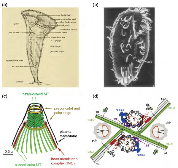
Cells are highly complex and orderly machines, with defined shapes and a startling variety of internal organizations. Complex geometry is a feature of both free-living unicellular organisms and cells inside multicellular animals. Where does the geometry of a cell come from? Many of the same questions that arise in developmental biology can also be asked of cells, but in most cases we do not know the answers. How much of cellular organization is dictated by global cell polarity cues as opposed to local interactions between cellular components? Does cellular structure persist across cell generations? What is the relationship between cell geometry and tissue organization? What ensures that intracellular structures are scaled to the overall size of the cell? Cell biology is only now beginning to come to grips with these questions.


Figures

Figure 1
Figure 1
Complexity in free-living eukaryotic cells. (a) The giant ciliate Stentor coeruleus, a classic system for studying cellular pattern formation using microsurgical methods [5]. Each cell can be up to 2 mm long and has a complex and highly asymmetrical morphology that can be faithfully regenerated following surgical manipulation. Image courtesy of Biodiversity Heritage Library. http://www.biodiversitylibrary.org[5]. (b) Ventral surface of Stylonychia [7] showing distinct classes of cirri arranged in highly asymmetrical patterns that are reproducible from cell to cell. Reprinted from Developmental Biology [7] with permission from Elsevier. (c) Apical complex (from which the apicomplexans take their name) of Toxoplasma cell [9] containing distinct sets of microtubule-based structures. (d) Basal apparatus of Chlamydomonas [11] showing the complex inter-relationship between the two mature basal bodies, the two daughter basal bodies formed prior to division, four microtubule-based rootlets, and several accessory fibers linking the rootlets to the basal bodies. These complex geometrical relations surrounding centrioles and basal bodies are likely a key source of local positional information. Reproduced with permission from Journal of Cell Science [11].
Figure 2
Figure 2
Complex intracellular structures in animal cells. (a) Stereocilia bundles [13]. (b) Retinal rod outer segment [14] showing well-ordered stacks of rhodopsin-containing membrane vesicles. Reproduced with permission from Journal of Neuroscience [66].
Figure 3
Figure 3
Global versus local information in cell morphogenesis as revealed by grafting in Stylonychia (modified from diagram in [46]). (a) Highly schematic view of paroral structures during normal development in Stylonychia, showing paroral membrane (PM; green) flanked by fronto-ventral-transverse (FVT) cirri (red). The oral primordium is shown as a grey disc. Other ciliary structures are not shown. Left and right (by convention given from the cell's perspective) are as indicated. (b) Grafting experiment of Grimes and L'Hernault [7]. (c, d) The cell was cut lengthwise, then the right half folded over (c), thus placing the former posterior region to the left of the former anterior region (d) with the join shown as a dotted line. (e) When PM and FVT cirri form, two sets are formed, one on the left and one on the right. The set on the left has inverted chirality because the structures have an anterior-posterior order consistent with the overall cell body axis after grafting, while the left-right ordering is consistent with the local position of these structures in the right half before cutting and folding.

Similar articles

Cited by

References

    1. Harrison SC, Brugge JS. Mechanistic biology in the next quarter century. Mol Biol Cell. 2010;21:3799–3800. doi: 10.1091/mbc.E10-05-0441. - DOI - PMC - PubMed
    1. Kirschner M, Gerhart J, Mitchison T. Molecular "vitalism". Cell. 2000;100:79–88. doi: 10.1016/S0092-8674(00)81685-2. - DOI - PubMed
    1. Shulman JM, St Johnston D. Pattern formation in single cells. Trends Cell Biol. 1999;9:M60–64. doi: 10.1016/S0962-8924(99)01680-3. - DOI - PubMed
    1. Aufderheide KJ, Frankel J, Williams NE. Formation and positioning of surface-related structures in protozoa. Microbiol Rev. 1980;44:252–302. - PMC - PubMed
    1. Tartar V. The Biology of Stentor. New York: Pergamon Press; 1961.

Publication types