. 2011 Dec;39(22):9695-704.
doi: 10.1093/nar/gkr692. Epub 2011 Aug 31.

A novel three-unit tRNA splicing endonuclease found in ultrasmall Archaea possesses broad substrate specificity

Affiliations

A novel three-unit tRNA splicing endonuclease found in ultrasmall Archaea possesses broad substrate specificity

Kosuke Fujishima et al. Nucleic Acids Res. 2011 Dec.

Abstract

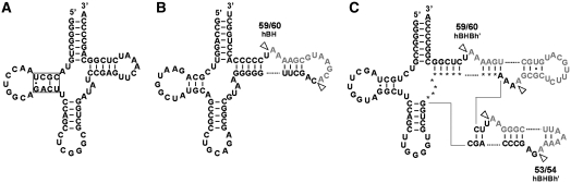
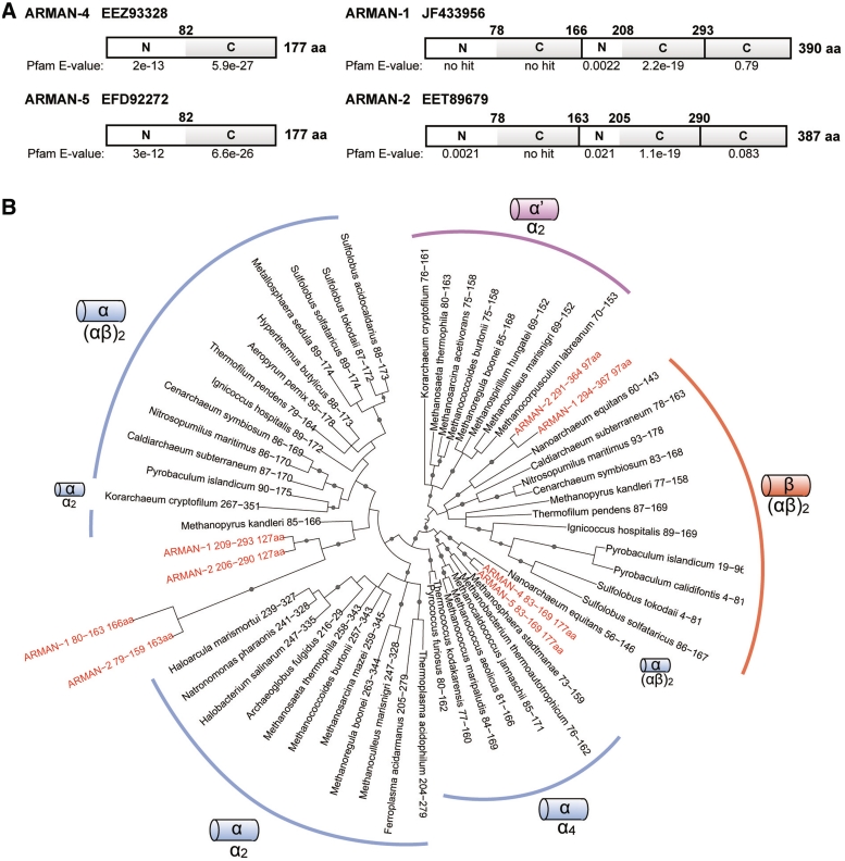
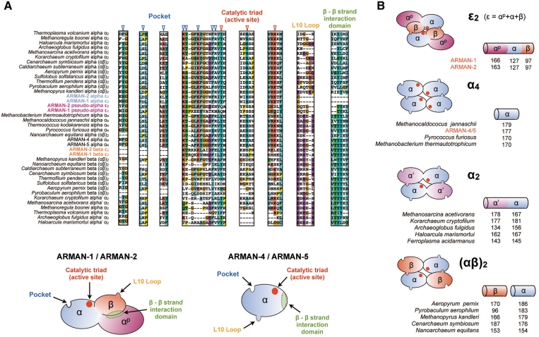
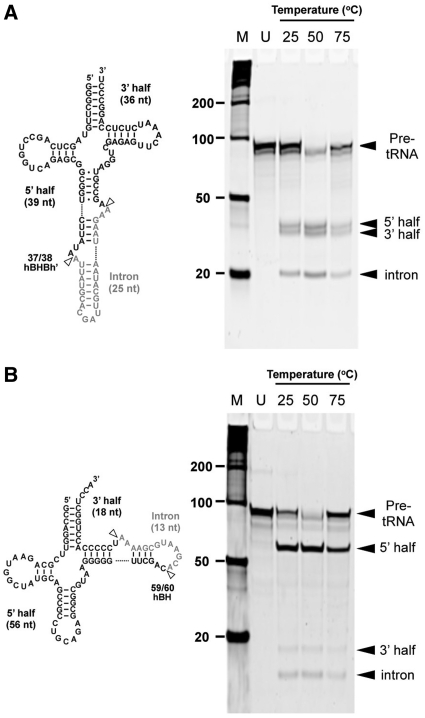
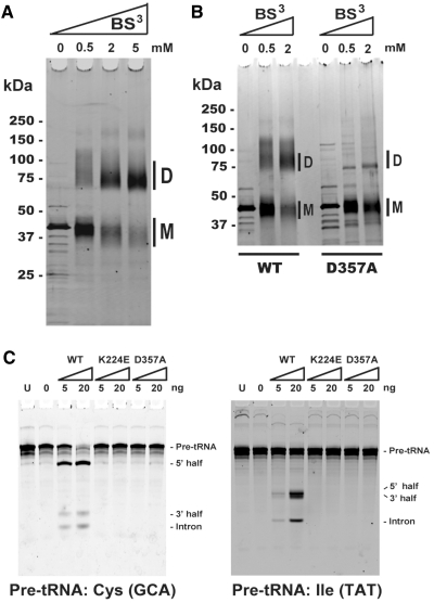
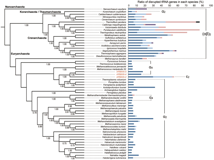
tRNA splicing endonucleases, essential enzymes found in Archaea and Eukaryotes, are involved in the processing of pre-tRNA molecules. In Archaea, three types of splicing endonuclease [homotetrameric: α(4), homodimeric: α(2), and heterotetrameric: (αβ)(2)] have been identified, each representing different substrate specificity during the tRNA intron cleavage. Here, we discovered a fourth type of archaeal tRNA splicing endonuclease (ε(2)) in the genome of the acidophilic archaeon Candidatus Micrarchaeum acidiphilum, referred to as ARMAN-2 and its closely related species, ARMAN-1. The enzyme consists of two duplicated catalytic units and one structural unit encoded on a single gene, representing a novel three-unit architecture. Homodimeric formation was confirmed by cross-linking assay, and site-directed mutagenesis determined that the conserved L10-pocket interaction between catalytic and structural unit is necessary for the assembly. A tRNA splicing assay reveal that ε(2) endonuclease cleaves both canonical and non-canonical bulge-helix-bulge motifs, similar to that of (αβ)(2) endonuclease. Unlike other ARMAN and Euryarchaeota, tRNAs found in ARMAN-2 are highly disrupted by introns at various positions, which again resemble the properties of archaeal species with (αβ)(2) endonuclease. Thus, the discovery of ε(2) endonuclease in an archaeon deeply branched within Euryarchaeota represents a new example of the coevolution of tRNA and their processing enzymes.


Figures

Figure 1.
Figure 1.
Schematics of the unique tRNAs found in ARMAN-2 genome. (A) tRNAPro (CGG) possessing a D-arm with a single Watson–Crick base pair (boxed). This feature is also found in synonymous tRNAPro(UGG). (B) Pre-tRNACys (GCA) with an intron located at the non-canonical position 59/60 forming relaxed BHB motif (hBH type). (C) Pre-tRNAPro (GGG) with introns located at two adjacent positions (53/54 and 59/60) within the T-loop forming a strict BHB motif (hBHBh′ type). Intron sequences are indicated by gray text.
Figure 2.
Figure 2.
Protein domain structure and phylogenetic positions of ARMAN splicing endonuclease units/subunits. (A) Domain structures of the putative splicing endonucleases found in ARMAN-1, -2, -4 and -5 (with there Genbank ID) are annotated by either N, tRNA intron endonuclease, N-terminal domain (Pfam family: PF02778) or C, tRNA intron endonuclease, catalytic C-terminal domain (Pfam family: PF01974), based on the Pfam domain search (19). Pfam E-value and amino acid (aa) position are indication for each domain. (B) Bayesian phylogenetic analysis of the catalytic C-terminal domain of total 59 archaeal splicing endonuclease units/subunits. The term ‘subunit’ is used for α4, (αβ)2 and ARMAN-4/5 endonucleases, whereas the term ‘unit’ is used for α2 and ARMAN-1/2 endonucleases that consist of duplicated or fused subunits. The catalytic α-units/subunits are phylogenetically distinguishable from the structural α′- and β-units/subunits. Gray dots on the branches indicate the posterior probability above 0.8.
Figure 3.
Figure 3.
Comparison of key amino acid residues and multimeric formation of ARMAN splicing endonuclease units/subunits. (A) Residues from four key conserved regions; catalytic triad, pocket, loop L10 and intrasubunit β–β interaction domain are compared among various archaeal splicing endonuclease units/subunits based on the structure-based sequence alignment shown in Supplementary Figure S2. Key residues are indicated on the cartoon model of ARMAN-2 ε2 endonuclease comprised of three units αp (purple), α (blue) and β (orange). Electrostatic interaction between positively charged pocket and negatively charged L10 loop is required for dimer/tetramer formation. Catalytic triad: tyrosine (Y), histidine (H) and lysine (K), is essential for RNA cleavage. Anti-parallel interaction of the two hydrophobic β–β interface is necessary for inter/intra-unit interaction such as α-β assembly in (αβ)2 endonuclease and α-α′ assembly in α2 endonuclease. (B) Proposed structural model and amino acid length of ε2 endonuclease units from ARMAN-1 and -2 are compared with those of other types of archaeal tRNA splicing endonucleases: homotetramer (α4), homodimer (α2) and heterotetramer [(αβ)2]. The tRNA splicing endonuclease found in ARMAN-4 and -5 belongs to α4 type.
Figure 4.
Figure 4.
In vitro splicing analysis of pre-tRNAs by ARMAN-2 tRNA splicing endonuclease. (A) Pre-tRNAIle (UAU) with 25 nt intron inserted at canonical position (37/38) forming a strict hBHBh′ motif and (B) Pre-tRNACys (GCA) with 13 nt intron located at non-canonical position (59/60) forming a relaxed hBH motif. Pre-tRNAs were incubated with the ARMAN-2 splicing endonuclease for 1 h at different temperatures. Cleaved products (black arrow) were analyzed by 8 M–urea 15% PAGE and stained with SYBR Green II. M, molecular marker; U, uncut product.
Figure 5.
Figure 5.
Enzyme assembly and splicing ability of WT and mutant proteins. The cross-linking assay was carried out by treating ∼500 ng of protein with 0, 0.5, 2 and 5 mM concentration of the crosslinker BS3 (Bis[sulfosuccinimidyl] suberate). The monomer and dimer are indicated as M and D. (A) Wild-type ARMAN-2 splicing endonuclease (B) WT and mutant protein with a single mutation at D357A in a side-by-side orientation. (C) Pre-tRNA splicing assay was carried out at 50°C for 1 h using WT and two mutant proteins (K224E and D357A) with different protein abundances (0, 5 and 20 ng) and 400 ng of pre-tRNA. Cleaved products were analyzed by 8 M–urea 15% PAGE and stained with SYBR Green II. U, uncut product.
Figure 6.
Figure 6.
Ratio of disrupted tRNA genes in archaeal evolution. Ratio of four types of disrupted tRNA genes; single intron located at canonical position 37/38 (blue), single intron located at non-canonical position (light blue), multiple introns (pink), split (purple) and permuted (black) are mapped on the SSU rRNA phylogenetic tree of 55 representative archaeal species (one species per genus) including three ARMAN lineages. Phylum names are indicated on the branch. Posterior probabilities above 0.75 are shown. Four types of splicing endonucleases [α4, α2, (αβ)2 and ε2] are denoted to the corresponding archaeal species.

References

    1. Marck C, Grosjean H. tRNomics: analysis of tRNA genes from 50 genomes of Eukarya, Archaea, and Bacteria reveals anticodon-sparing strategies and domain-specific features. RNA. 2002;8:1189–1232. - PMC - PubMed
    1. Sugahara J, Kikuta K, Fujishima K, Yachie N, Tomita M, Kanai A. Comprehensive analysis of archaeal tRNA genes reveals rapid increase of tRNA introns in the order thermoproteales. Mol. Biol. Evol. 2008;25:2709–2716. - PubMed
    1. Calvin K, Li H. RNA-splicing endonuclease structure and function. Cell Mol. Life Sci. 2008;65:1176–1185. - PMC - PubMed
    1. Li H, Trotta CR, Abelson J. Crystal structure and evolution of a transfer RNA splicing enzyme. Science. 1998;280:279–284. - PubMed
    1. Tocchini-Valentini GD, Fruscoloni P, Tocchini-Valentini GP. Structure, function, and evolution of the tRNA endonucleases of Archaea: an example of subfunctionalization. Proc. Natl Acad. Sci. USA. 2005;102:8933–8938. - PMC - PubMed

Publication types

Associated data