. 2011 Nov 11;286(45):39528-36.
doi: 10.1074/jbc.M111.278812. Epub 2011 Aug 31.

Crystal structure of NALP3 protein pyrin domain (PYD) and its implications in inflammasome assembly

Affiliations

Crystal structure of NALP3 protein pyrin domain (PYD) and its implications in inflammasome assembly

Ju Young Bae et al. J Biol Chem. .

Abstract

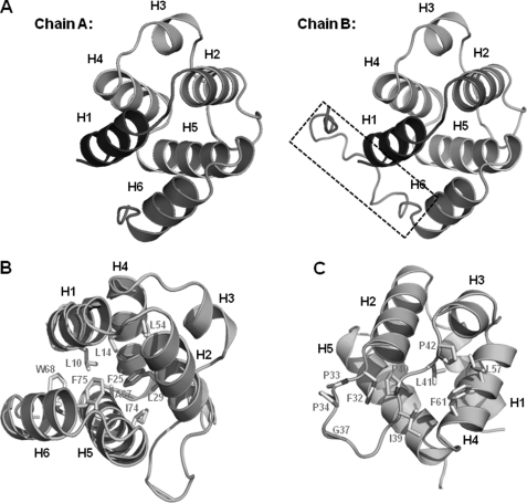
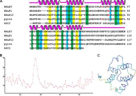
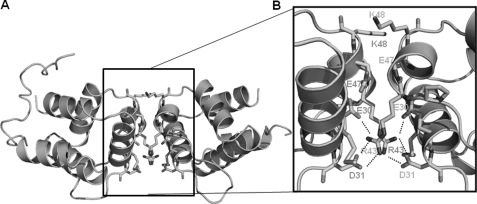
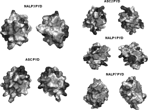
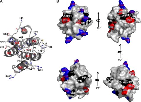
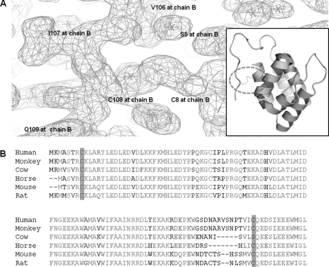
NALP3 inflammasome, composed of the three proteins NALP3, ASC, and Caspase-1, is a macromolecular complex responsible for the innate immune response against infection with bacterial and viral pathogens. Formation of the inflammasome can lead to the activation of inflammatory caspases, such as Caspase-1, which then activate pro-inflammatory cytokines by proteolytic cleavage. The assembly of the NALP3 inflammasome depends on the protein-interacting domain known as the death domain superfamily. NALP3 inflammasome is assembled via a pyrin domain (PYD)/PYD interaction between ASC and NALP3 and a caspase recruitment domain/caspase recruitment domain interaction between ASC and Caspase-1. As a first step toward elucidating the molecular mechanisms of inflammatory caspase activation by formation of inflammasome, we report the crystal structure of the PYD from NALP3 at 1.7-Å resolution. Although NALP3 PYD has the canonical six-helical bundle structural fold similar to other PYDs, the high resolution structure reveals the possible biologically important homodimeric interface and the dynamic properties of the fold. Comparison with other PYD structures shows both similarities and differences that may be functionally relevant. Structural and sequence analyses further implicate conserved surface residues in NALP3 PYD for ASC interaction and inflammasome assembly. The most interesting aspect of the structure was the unexpected disulfide bond between Cys-8 and Cys-108, which might be important for regulation of the activity of NALP3 by redox potential.


Figures

FIGURE 1.
FIGURE 1.
Crystal structure of NALP3 PYD. A, ribbon diagram of NALP3 PYD. Chain A and chain B are shown separately. Helices are labeled. Extra structure of C terminus on chain B is shown by a black dashed box. B, conserved central hydrophobic core. Hydrophobic residues essential for the formation of this core that stabilizes H1 to H6 (except H3) are shown as a gray stick. C, second hydrophobic patch. Helices are labeled, and residues involved in the formation of exposed second hydrophobic patch are shown as a gray stick.
FIGURE 2.
FIGURE 2.
Structural based sequence alignment and B-factor distribution. A, structure-based sequence alignment of NALP3 PYD with other ASC-binding proteins. Secondary structures (helices H1 to H6) are shown above the sequences. Residues at the hydrophobic core are shaded in yellow; conserved exposed hydrophobic and charged residues are shaded in green and cyan, respectively. Residues involved in the interaction between chain A and chain B are colored in red. B, B-factor distribution. Whole residue B-factors are plotted. The x and y axes indicate residue number and B-factor, respectively. C, B-factor distribution on the structure. Warm and cold colors indicate high and low B-factors, respectively.
FIGURE 3.
FIGURE 3.
Dimeric interface of the structure of NALP3 PYD. A, dimeric structure of NALP3 PYD. B, close-up view of the interacting residues in the interface between two dimers. Helices are labeled, and residues involved in the contact are shown as sticks. Salt bridges formed between Arg-43 from one chain and Glu-30 and Asp-31 from counterparts are shown as dashed lines.
FIGURE 4.
FIGURE 4.
Superposition of NALP3 PYD with their structural homologues. NALP3 PYD and four structural homologues are superimposed (green, NALP3 PYD; cyan, ASC PYD; magenta, ASC2 PYD; yellow, NALP7 PYD; blue, NALP1 PYD). Pairwise structural comparisons are also performed. NALP3 PYD is colored in green, and each counterpart is colored in cyan for ASC PYD, magenta for ASC2 PYD, yellow for NALP7 PYD, and blue for NALP1 PYD.
FIGURE 5.
FIGURE 5.
Electrostatic surface representation of NALP3 PYD and their structural homologues. The left side of the NALP3 PYD surface and the remaining PYDs are shown in the same orientations as in Fig. 3. The opposite side of NALP3 PYD and the other PYDs are rotated by 180° along the vertical axis (y).
FIGURE 6.
FIGURE 6.
Mapping of conserved exposed residues onto the NALP3 PYD. A, ribbon diagram of NALP3 PYD with conserved surface residues. B, molecular surface representation of NALP3 PYD with conserved exposed residues colored and labeled. Positively charged residues and negatively charged residues are colored in blue and red, respectively. Conserved exposed hydrophobic residues are colored in black. Four orientations of NALP3 PYD surface are shown.
FIGURE 7.
FIGURE 7.
Disulfide bond between Cys-8 and Cys-108 and conserved cysteine residue cross-species. A, electron density map is shown and disulfide bond is shown as sticks. Disulfide bond on the ribbon diagram is also shown in the boxed figure. The dotted circle indicates the position of the disulfide bond. B, conserved cysteine residues that are involved in the disulfide bond. The conserved cysteine residue cross-species are highlighted.

Similar articles

Cited by

References

    1. Schroder K., Tschopp J. (2010) Cell 140, 821–832 - PubMed
    1. Eisenbarth S. C., Flavell R. A. (2009) EMBO Mol. Med. 1, 92–98 - PMC - PubMed
    1. Martinon F., Burns K., Tschopp J. (2002) Mol. Cell 10, 417–426 - PubMed
    1. Faustin B., Lartigue L., Bruey J. M., Luciano F., Sergienko E., Bailly-Maitre B., Volkmann N., Hanein D., Rouiller I., Reed J. C. (2007) Mol. Cell 25, 713–724 - PubMed
    1. Agostini L., Martinon F., Burns K., McDermott M. F., Hawkins P. N., Tschopp J. (2004) Immunity 20, 319–325 - PubMed

Publication types

Associated data