. 2011 Oct 21;286(42):36992-7001.
doi: 10.1074/jbc.M111.263780. Epub 2011 Aug 31.

Identification of novel Ssl0352 protein (NdhS), essential for efficient operation of cyclic electron transport around photosystem I, in NADPH:plastoquinone oxidoreductase (NDH-1) complexes of Synechocystis sp. PCC 6803

Affiliations

Identification of novel Ssl0352 protein (NdhS), essential for efficient operation of cyclic electron transport around photosystem I, in NADPH:plastoquinone oxidoreductase (NDH-1) complexes of Synechocystis sp. PCC 6803

Natalia Battchikova et al. J Biol Chem. .

Erratum in

  • J Biol Chem. 2012 Mar 9;287(11):8660

Abstract

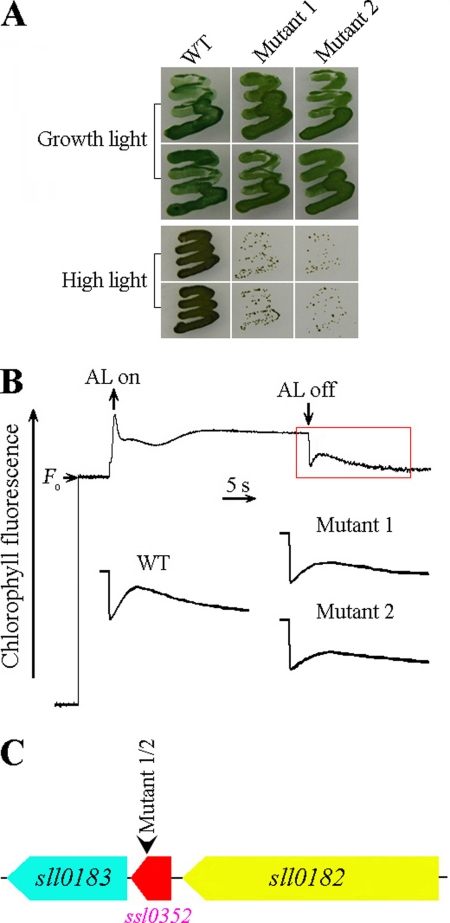
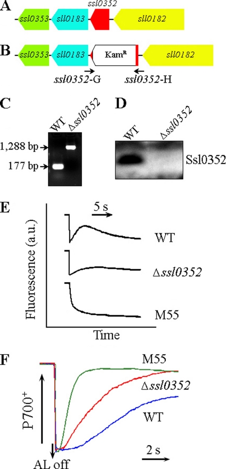
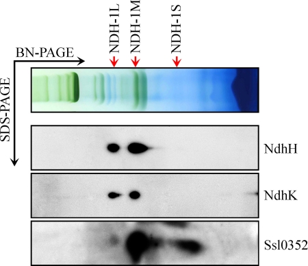
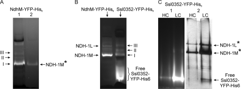
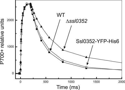
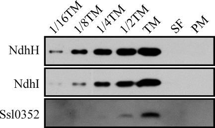
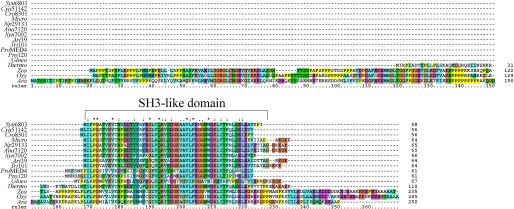
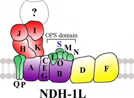
Cyanobacterial NADPH:plastoquinone oxidoreductase, or type I NAD(P)H dehydrogenase, or the NDH-1 complex is involved in plastoquinone reduction and cyclic electron transfer (CET) around photosystem I. CET, in turn, produces extra ATP for cell metabolism particularly under stressful conditions. Despite significant achievements in the study of cyanobacterial NDH-1 complexes during the past few years, the entire subunit composition still remains elusive. To identify missing subunits, we screened a transposon-tagged library of Synechocystis 6803 cells grown under high light. Two NDH-1-mediated CET (NDH-CET)-defective mutants were tagged in the same ssl0352 gene encoding a short unknown protein. To clarify the function of Ssl0352, the ssl0352 deletion mutant and another mutant with Ssl0352 fused to yellow fluorescent protein (YFP) and the His(6) tag were constructed. Immunoblotting, mass spectrometry, and confocal microscopy analyses revealed that the Ssl0352 protein resides in the thylakoid membrane and associates with the NDH-1L and NDH-1M complexes. We conclude that Ssl0352 is a novel subunit of cyanobacterial NDH-1 complexes and designate it NdhS. Deletion of the ssl0352 gene considerably impaired the NDH-CET activity and also retarded cell growth under high light conditions, indicating that NdhS is essential for efficient operation of NDH-CET. However, the assembly of the NDH-1L and NDH-1M complexes and their content in the cells were not affected in the mutant. NdhS contains a Src homology 3-like domain and might be involved in interaction of the NDH-1 complex with an electron donor.


Figures

FIGURE 1.
FIGURE 1.
High light strategy for screening NDH-CET-defective mutants in transposon-tagged mutant populations of Synechocystis 6803. A, growth phenotype of WT and mutants under growth light (40 μmol of photons m−2 s−1) and high light (200 μmol of photons m−2 s−1). B, monitoring of NDH-CET activity using chlorophyll fluorescence analysis. The top curve indicates a typical trace of chlorophyll fluorescence in the WT Synechocystis 6803. The chlorophyll a concentration was adjusted to 10 μg ml−1 before measurement. Cells were exposed to AL (620 nm; 45 μmol of photons m−2 s−1) for 30 s. AL was turned off, and the subsequent change in the chlorophyll fluorescence level was monitored as an indicator of NDH-CET activity. C, PCR analysis of the transposon insertion site using the primers listed in supplemental Table 1.
FIGURE 2.
FIGURE 2.
ssl0352 gene deletion mutation and its effect on NDH-CET. A, WT locus of the ssl0352 gene. B, ΔSsl0352 mutant locus in which the majority of the coding region of the ssl0352 gene has been replaced by a kanamycin resistance marker (KamR). C, PCR segregation analysis of the Δssl0352 mutant using the ssl0352-G and ssl0352-H primers (supplemental Table 1) whose positions are labeled in B. D, immunoblotting inactivation analysis of the ssl0352 gene in Synechocystis 6803 using the specific Ssl0352 antibody. E, monitoring of NDH-CET activity by chlorophyll fluorescence. 2-day-old cells were exposed to AL (45 μmol of photons m−2 s−1) for 30 s. After illumination, the subsequent transient change in chlorophyll fluorescence was monitored as an indicator of NDH-CET activity. a.u., arbitrary units. F, redox kinetics of P700 after termination of AL illumination (800 μmol of photons m−2 s−1 for 30 s) under a background of FR. The cells were illuminated by AL supplemented with FR to store electrons in the stromal pool. After termination of AL illumination, P700+ was transiently reduced by electrons from the plastoquinone pool; thereafter, P700 was reoxidized by background FR. The redox kinetics of P700 was recorded. The P700+ levels were standardized by their maximum levels attained by exposure to FR.
FIGURE 3.
FIGURE 3.
Identification of Ssl0352 protein in NDH-1 complexes by Western blotting. NDH-1 complexes isolated from the thylakoid membrane of WT Synechocystis cells were separated by BN-PAGE and further subjected to two-dimensional SDS-PAGE. The proteins were probed with specific antibodies against NdhH, -K, and Ssl0352. The positions of the NDH-1L, NDH-1M, and NDH-1S complexes, respectively, in WT are indicated by arrows.
FIGURE 4.
FIGURE 4.
Discovery of Ssl0352 protein in NDH-1 complexes containing YFP-His6 tag-fused subunits and purified by Ni2+ affinity chromatography. A, fluorescent protein complexes from the NdhM-YFP-His6 mutant before (A) and after (B) chromatography on a Ni2+ column (4.5–8% CN-PAGE). B, fluorescent protein complexes in the Ssl0352-YFP-His6 mutant (4.5–12.5% CN-PAGE). C, CN-PAGE (4.5–12.5%) analysis of the protein complexes from the Ssl0352-YFP-His6 mutant that were retained on Ni2+ resin. HC and LC, high CO2 and low CO2 growth conditions, respectively. Proteins were visualized by fluorescence (panel 1) and by silver staining (panel 2). YFP fluorescence was detected as described under “Experimental Procedures.” The NDH-1 complexes verified by the MS/MS analysis are shown by a star. Bands I, II, and III correspond to NDH-1M, NDH-1I, and NDH-1L, respectively.
FIGURE 5.
FIGURE 5.
P700+ oxidoreduction kinetics in WT (closed squares), Δssl0352 mutant (closed triangles), and Ssl0352-YFP-His6 mutant (open circles).
FIGURE 6.
FIGURE 6.
Localization of Ssl0352 protein. The purified thylakoid (TM) and plasma (PM) membrane fractions were obtained by sucrose density fractionation followed by the two-phase partitioning system composed of dextran and polyethylene glycol. Serial dilutions of thylakoid membrane are indicated. SF, the fraction of soluble proteins. Immunoblotting was performed with antibodies against NdhH, NdhI, and Ssl0352.
FIGURE 7.
FIGURE 7.
Sequence alignment of Ssl0352 protein of Synechocystis 6803 and its homologues from other species. The sequence of the Ssl0352 protein from Synechocystis sp. PCC 6803 (Syn6803) was compared with related sequences from Cyanothece sp. ATCC 51142 (Cya51142), Crocosphaera watsonii WH8501 (Cro8501), Microcystis aeruginosa NIES-843 (Micro), Nostoc punctiforme ATCC 29133 (Np29133), Anabaena sp. PCC 7120 (Ana7120), Synechococcus sp. PCC 7002 (Syn7002), Arthrospira platensis NIES-39 (Art39), Trichodesmium erythraeum IMS101 (Tri101), Prochlorococcus marinus MED4 (ProMED4), Prochlorococcus marinus SS120 (Pro120), Gloeobacter violaceus PCC 7421 (Gloeo), T. elongatus BP-1 (Thermo), Zea mays (Zea), Oryza sativa (Ory), and A. thaliana (Ara). Domain analysis was performed by the pfam software. The sequences were aligned using ClustalX 1.83. Asterisks indicate identical amino acids; colons and dots indicate conserved amino acid substitutions.
FIGURE 8.
FIGURE 8.
Model of cyanobacterial NDH-1L complex. An oxygenic photosynthesis-specific (OPS) domain is indicated, and the new NDH-1 subunit NdhS is included in this oxygenic photosynthesis-specific domain. The unknown subunits responsible for NADPH oxidation are indicated by the question mark.

References

    1. Ogawa T. (1991) Proc. Natl. Acad. Sci. U.S.A. 88, 4275–4279 - PMC - PubMed
    1. Mi H., Endo T., Schreiber U., Ogawa T., Asada K. (1992) Plant Cell Physiol. 33, 1233–1237
    1. Ohkawa H., Pakrasi H. B., Ogawa T. (2000) J. Biol. Chem. 275, 31630–31634 - PubMed
    1. Friedrich T., Steinmüller K., Weiss H. (1995) FEBS Lett. 367, 107–111 - PubMed
    1. Friedrich T., Scheide D. (2000) FEBS Lett. 479, 1–5 - PubMed

Publication types

MeSH terms

Substances

LinkOut - more resources