. 2011 Nov;85(22):11581-7.
doi: 10.1128/JVI.05662-11. Epub 2011 Aug 31.

Tissue tropism of swine influenza viruses and reassortants in ex vivo cultures of the human respiratory tract and conjunctiva

Affiliations

Tissue tropism of swine influenza viruses and reassortants in ex vivo cultures of the human respiratory tract and conjunctiva

Renee W Y Chan et al. J Virol. 2011 Nov.

Abstract

The 2009 pandemic influenza H1N1 (H1N1pdm) virus was generated by reassortment of swine influenza viruses of different lineages. This was the first influenza pandemic to emerge in over 4 decades and the first to occur after the realization that influenza pandemics arise from influenza viruses of animals. In order to understand the biological determinants of pandemic emergence, it is relevant to compare the tropism of different lineages of swine influenza viruses and reassortants derived from them with that of 2009 pandemic H1N1 (H1N1pdm) and seasonal influenza H1N1 viruses in ex vivo cultures of the human nasopharynx, bronchus, alveoli, and conjunctiva. We hypothesized that virus which can transmit efficiently between humans replicated well in the human upper airways. As previously reported, H1N1pdm and seasonal H1N1 viruses replicated efficiently in the nasopharyngeal, bronchial, and alveolar epithelium. In contrast, representative viruses from the classical swine (CS) (H1N1) lineage could not infect human respiratory epithelium; Eurasian avian-like swine (EA) (H1N1) viruses only infected alveolar epithelium and North American triple-reassortant (TRIG) viruses only infected the bronchial epithelium albeit inefficiently. Interestingly, a naturally occurring triple-reassortant swine virus, A/SW/HK/915/04 (H1N2), with a matrix gene segment of EA swine derivation (i.e., differing from H1N1pdm only in lacking a neuraminidase [NA] gene of EA derivation) readily infected and replicated in human nasopharyngeal and bronchial epithelia but not in the lung. A recombinant sw915 with the NA from H1N1pdm retained its tropism for the bronchus and acquired additional replication competence for alveolar epithelium. In contrast to H1N1pdm, none of the swine viruses tested nor seasonal H1N1 had tropism in human conjunctiva. Recombinant viruses generated by swapping the surface proteins (hemagglutinin and NA) of H1N1pdm and seasonal H1N1 virus demonstrated that these two gene segments together are key determinants of conjunctival tropism. Overall, these findings suggest that ex vivo cultures of the human respiratory tract provide a useful biological model for assessing the human health risk of swine influenza viruses.


Figures
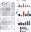
Fig. 1.
Fig. 1.
Expression of influenza virus nucleoprotein (reddish brown) in the upper, conducting, and lower respiratory tract (A, D, G, J, M, and P), in the nasopharynx (B, E, H, K, N, and Q), and in the bronchi and lungs (C, F, I, L, O, and R) infected with influenza virus A/HK/415742/09 (H1N1pdm), A/SW/HK/4167/99 (H1N1), A/SW/AR/2976/02 (H1N2), A/SW/HK/915/04 (H1N2), and A/SW/HK/201/10 (H1N1) at 48 h postinfection (hpi). Viral replication kinetics in ex vivo cultures of nasopharynx (S), bronchi (T), and lung biopsy specimens (U) infected with 106 TCID50 of influenza viruses/ml by virus titration at 37°C were also evaluated. The chart shows the means and standard errors of the means of the virus titer pooled from at least three independent experiments. Horizontal dotted line denotes the detection limit of the viral titration assay. Colored asterisks indicate the statistically significant increases in viral yield compared to 1 hpi, and black asterisks indicate statistically significant differences between HK415742/H1N1pdm and sw201/H1N1 in figure U. **, P < 0.005.
Fig. 2.
Fig. 2.
Expression of influenza virus nucleoprotein (reddish brown) in the bronchus (A) and in lung biopsy specimens (B) by recombinant influenza A virus of rg sw915/H1N2 with the insertion of NA from HK415742/H1N1pdm influenza virus at 48 hpi. (C) Viral replication kinetics in ex vivo cultures of bronchi and lungs infected with 106 TCID50 of HK415742/H1N1pdm, sw915/H1N2, and rg sw915/H1N2 with the insertion of NA from HK415742/H1N1pdm influenza virus by virus titration at 37°C. Colored asterisks indicate statistically significant increases in the viral yield compared to 1 hpi, and black asterisks represent statistically significant differences between HK415742/H1N1pdm and rg sw915/H1N2 with the insertion of NA from HK415742/H1N1pdm. ***, P < 0.0005. The chart showed the means and the standard errors of the means of the virus titer pooled from three independent experiments. Asterisks indicate statistically significant increases in viral yield compare to 1 hpi: *, P < 0.05; **, P < 0.005; and ***, P < 0.0005.
Fig. 3.
Fig. 3.
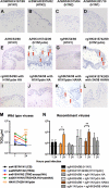
Ex vivo organ cultures of conjunctiva infected with wild-type influenza A/SW/HK/4167/99 (H1N1) (A), A/SW/AR/2976/02 (H1N2) (B), A/SW/HK/915/04 (H1N2) (C), A/SW/HK/201/10 (H1N1) (D), A/HK/54/98 (H1N1) (E), and A/HK/415742/09 (H1N1pdm) (F) viruses and the recombinant influenza viruses rg/HK/54/98 (H1N1) (G), rg/HK/415742/09 (H1N1pdm) (F), rg/HK/54/98 with the HA of A/HK/415742/09 (H1N1pdm) (I), rg/HK/54/98 with the NA of A/HK/415742/09 (H1N1pdm) (J), rg/HK/54/98 with the HANA of A/HK/415742/09 (H1N1pdm) (K), and rg/HK/415742/09 (H1N1pdm) with the HANA of A/HK/54/98 (H1N1) (L) at 24 hpi and stained for influenza virus A nucleoprotein in reddish brown, as indicated by arrows. (M and N) Wild-type (M) and recombinant (N) influenza virus yield at 24 hpi after infection with 106 TCID50 of the virus/ml at 33°C. The chart shows the means and the standard errors of the virus titers pooled from three independent experiments. Horizontal dotted line denotes detection limit of the viral titration. *, P < 0.05; **, P < 0.005; ***, P < 0.0005.

References

    1. Chan M. C., et al. 2010. Tropism and innate host responses of the 2009 pandemic H1N1 influenza virus in ex vivo and in vitro cultures of human conjunctiva and respiratory tract. Am. J. Pathol. 176: 1828–1840 - PMC - PubMed
    1. Dawood F. S., et al. 2009. Emergence of a novel swine-origin influenza A (H1N1) virus in humans. N. Engl. J. Med. 360: 2605–2615 - PubMed
    1. Garten R. J., et al. 2009. Antigenic and genetic characteristics of swine-origin 2009 A(H1N1) influenza viruses circulating in humans. Science 325: 197–201 - PMC - PubMed
    1. Ilyushina N. A., et al. 2010. Adaptation of pandemic H1N1 influenza viruses in mice. J. Virol. 84: 8607–8616 - PMC - PubMed
    1. Lee S. M., et al. 2010. Systems-level comparison of host responses induced by pandemic and seasonal influenza A H1N1 viruses in primary human type I-like alveolar epithelial cells in vitro. Respir. Res. 11: 147. - PMC - PubMed

Publication types

MeSH terms