

Direct sequencing and characterization of a clinical isolate of Epstein-Barr virus from nasopharyngeal carcinoma tissue by using next-generation sequencing technology
- PMID: 21880770
- PMCID: PMC3194977
- DOI: 10.1128/JVI.00823-11
Direct sequencing and characterization of a clinical isolate of Epstein-Barr virus from nasopharyngeal carcinoma tissue by using next-generation sequencing technology
Abstract
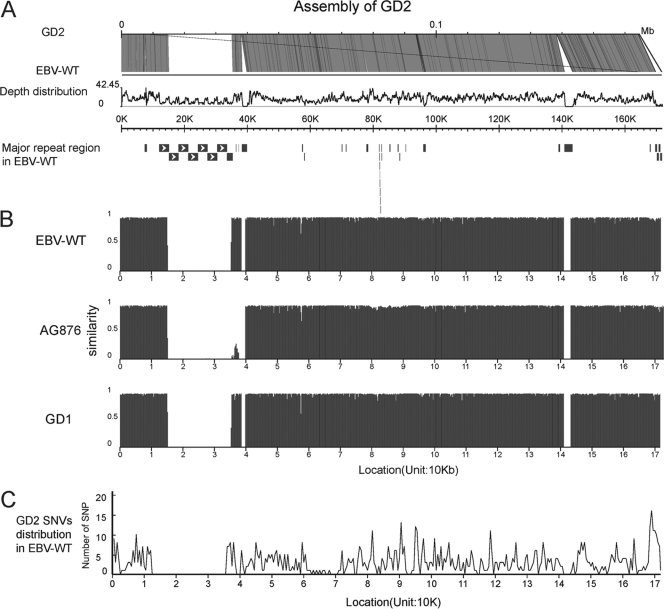
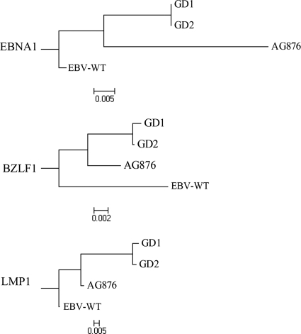
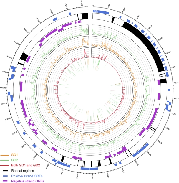
Epstein-Barr virus (EBV)-encoded molecules have been detected in the tumor tissues of several cancers, including nasopharyngeal carcinoma (NPC), suggesting that EBV plays an important role in tumorigenesis. However, the nature of EBV with respect to genome width in vivo and whether EBV undergoes clonal expansion in the tumor tissues are still poorly understood. In this study, next-generation sequencing (NGS) was used to sequence DNA extracted directly from the tumor tissue of a patient with NPC. Apart from the human sequences, a clinically isolated EBV genome 164.7 kb in size was successfully assembled and named GD2 (GenBank accession number HQ020558). Sequence and phylogenetic analyses showed that GD2 was closely related to GD1, a previously assembled variant derived from a patient with NPC. GD2 contains the most prevalent EBV variants reported in Cantonese patients with NPC, suggesting that it might be the prevalent strain in this population. Furthermore, GD2 could be grouped into a single subtype according to common classification criteria and contains only 6 heterozygous point mutations, suggesting the monoclonal expansion of GD2 in NPC. This study represents the first genome-wide analysis of a clinical isolate of EBV directly extracted from NPC tissue. Our study reveals that NGS allows the characterization of genome-wide variations of EBV in clinical tumors and provides evidence of monoclonal expansion of EBV in vivo. The pipeline could also be applied to the study of other pathogen-related malignancies. With additional NGS studies of NPC, it might be possible to uncover the potential causative EBV variant involved in NPC.
Figures

References
-
- Adldinger H. K., Delius H., Freese U. K., Clarke J., Bornkamm G. W. 1985. A putative transforming gene of Jijoye virus differs from that of Epstein-Barr virus prototypes. Virology 141:221–234 - PubMed
-
- Ansorge W. J. 2009. Next-generation DNA sequencing techniques. N. Biotechnol. 25:195–203 - PubMed
-
- Baer R., et al. 1984. DNA sequence and expression of the B95-8 Epstein-Barr virus genome. Nature 310:207–211 - PubMed
-
- Busson P., Keryer C., Ooka T., Corbex M. 2004. EBV-associated nasopharyngeal carcinomas: from epidemiology to virus-targeting strategies. Trends Microbiol. 12:356–360 - PubMed
-
- Chang E. T., Adami H. O. 2006. The enigmatic epidemiology of nasopharyngeal carcinoma. Cancer Epidemiol. Biomarkers Prev. 15:1765–1777 - PubMed
Publication types
MeSH terms
Substances
Associated data
- Actions
LinkOut - more resources
Full Text Sources
Other Literature Sources
