. 2011 Dec;301(6):F1358-66.
doi: 10.1152/ajprenal.00332.2011. Epub 2011 Aug 31.

Monocyte/macrophage chemokine receptor CCR2 mediates diabetic renal injury

Affiliations

Monocyte/macrophage chemokine receptor CCR2 mediates diabetic renal injury

Alaa S Awad et al. Am J Physiol Renal Physiol. 2011 Dec.

Abstract

Monocyte/macrophage recruitment correlates strongly with the progression of renal impairment in diabetic nephropathy (DN). C-C chemokine receptor (CCR)2 regulates monocyte/macrophage migration into injured tissues. However, the direct role of CCR2-mediated monocyte/macrophage recruitment in diabetic kidney disease remains unclear. We report that pharmacological blockade or genetic deficiency of CCR2 confers kidney protection in Ins2(Akita) and streptozotocin (STZ)-induced diabetic kidney disease. Blocking CCR2 using the selective CCR2 antagonist RS504393 for 12 wk in Ins2(Akita) mice significantly attenuated albuminuria, the increase in blood urea nitrogen and plasma creatinine, histological changes, and glomerular macrophage recruitment compared with vehicle. Furthermore, mice lacking CCR2 (CCR2(-/-)) mimicked CCR2 blockade by reducing albuminuria and displaying less fibronectin mRNA expression and inflammatory cytokine production compared with CCR2(+/+) mice, despite comparable blood glucose levels. Bone marrow-derived monocytes from CCR2(+/+) or CCR2(-/-) mice adoptively transferred into CCR2(-/-) mice reversed the renal tissue-protective effect in diabetic CCR2(-/-) mice as evaluated by increased urinary albumin excretion and kidney macrophage recruitment, indicating that CCR2 is not required for monocyte migration from the circulation into diabetic kidneys. These findings provide evidence that CCR2 is necessary for monocyte/macrophage-induced diabetic renal injury and suggest that blocking CCR2 could be a novel therapeutic approach in the treatment of DN.


Figures

Fig. 1.
Fig. 1.
Effects of a C-C chemokine receptor 2 (CCR2) antagonist on urinary albumin excretion (UAE) in Ins2Akita mice. Ins2Akita and their wild-type littermates were treated with a CCR2 antagonist (RS504393; 2 mg·kg−1·day−1) or vehicle via an osmotic minipump for 12 wk. Urine was collected for measurement of UAE before treatment (6 wk of age) and after treatment (18 wk of age). Open bars, vehicle-treated groups; filled bars, CCR2 antagonist-treated groups. Values are means ± SE. *P < 0.05, **P < 0.001 vs. control. #P < 0.005 vs. Ins2Akita vehicle-treated.
Fig. 2.
Fig. 2.
Effect of CCR2 antagonist on histological changes in Ins2Akita mice. Sections were stained with periodic acid-Schiff (PAS), and all glomeruli were examined at ×25. They were then graded at ×40. Images were taken with ×100 (oil) objective with a total magnification of ×1,000. Images are representative of 5–8 mice/group at 18 wk of age in control mice treated with vehicle (A), control mice treated with CCR2 antagonist (B), Ins2Akita mice treated with vehicle (C), and Ins2Akita mice treated with CCR2 antagonist (D).
Fig. 3.
Fig. 3.
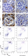
Effect of CCR2 antagonist on macrophage recruitment in Ins2Akita mice. A: immunohistochemical staining for Mac-2-positive macrophages in glomeruli at 18 wk of age in control mice treated with vehicle, control mice treated with CCR2 antagonist, Ins2Akita mice treated with vehicle, and Ins2Akita mice treated with CCR2 antagonist. Images are representative of 20 fields from 5–8 mice/group. B: experimental groups were as described in A. Kidneys were harvested at 18 wk of age and processed for fluorescence-activated cell sorting (FACS) analysis as described in materials and methods. Macrophages were identified as CD11bhighF4/80low. Graphs show representative contour plots. Values are means ± SE expressed as numbers of CD11bhighF4/80low macrophages/g kidney tissue; n = 5–8 mice/group.
Fig. 4.
Fig. 4.
Effect of CCR2 deletion on UAE in diabetes. To determine whether CCR2 contributes to diabetic renal injury, urine was collected for measurement of UAE in CCR2+/+ and CCR2−/− mice (control and diabetic groups; n = 8/group) at 6 wk after injection of streptozotocin (STZ) or vehicle. Open bars, control groups; filled bars, diabetic groups. Values are means ± SE. *P < 0.0001 vs. control CCR2+/+. #P < 0.0001 vs. diabetic CCR2+/+.
Fig. 5.
Fig. 5.
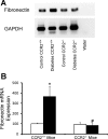
Effect of CCR2 deletion on fibronectin mRNA expression in diabetes. A and B: RT-PCR was performed on whole mouse kidney total RNA isolated after 6 wk of the study. A: gel analysis of PCR products. B: expression of fibronectin mRNA was normalized to GAPDH, and data were calculated as expression relative to control. Open bars, control groups; filled bars, diabetic groups. Values are means ± SE. *P < 0.05 vs. control CCR2+/+. #P < 0.005 vs. diabetic CCR2+/+.
Fig. 6.
Fig. 6.
Effect of diabetes on urinary TNF-α levels in CCR2−/− mice. Twenty-four-hour urine collections were obtained for measurement of TNF-α in CCR2+/+ and CCR2−/− mice (control and diabetic groups; n = 8/group). Open bars, control groups; filled bars, diabetic groups. Values are means ± SE. *P < 0.001 vs. CCR2+/+ control. #P < 0.001 vs. CCR2+/+ diabetes.
Fig. 7.
Fig. 7.
Renal tissue-protective effect in CCR2−/− mice is mediated via reducing monocyte/macrophage recruitment in diabetic nephropathy. To determine the mechanism of renal tissue-protective effects of CCR2−/− mice in DN, we adoptively transferred bone-marrow derived monocytes isolated by negative selection from CCR2+/+ or CCR2−/− mice into diabetic CCR2−/− mice (5 × 10⁶ cells at days 3 and 10 after STZ-induced diabetes) and followed them for the 6-wk study period. Urine collections were obtained for measurement of UAE (n = 5–7/group). Values are means ± SE. *P < 0.05 to normal CCR2+/+ group. #P < 0.01 to normal CCR2−/− group. +P < 0.05, ++P < 0.01 to diabetes CCR2−/− group.
Fig. 8.
Fig. 8.
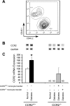
Efficiency of monocyte transfer in diabetic kidneys. A: monocytes were isolated from bone marrow of B6.SJL-Ptprca Pep3b/BoyJ mice (which carry CD45.1 antigen) by negative selection and injected (5 × 10⁶ cells at days 3 and 10 after STZ-induced diabetes) into C57BL/6 mice (which express CD45.2 antigen in leukocytes; n = 9). After 6 wk, kidneys were harvested and processed for FACS. Macrophages were identified as CD11bhighF4/80low. Graph shows representative contour plot of CD11bhighF4/80low/CD45.1+ (donor macrophages) against CD11bhighF4/80low/CD45.2+ (recipient macrophages). B and C: RT-PCR was performed on whole mouse kidney total RNA isolated after 6 wk of the study. B: gel analysis of PCR products. C: expression of CCR2 mRNA was normalized to GAPDH, and data were calculated as expression relative to control. Values are means ± SE. ND, nondetectable. *P < 0.05 vs. normal CCR2+/+.
Fig. 9.
Fig. 9.
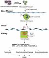
Proposed scheme for the role of CCR2 in monocyte/macrophage-induced kidney injury in diabetes. Mobilization of monocytes from the bone marrow to the blood is CCR2 dependent under normal homeostatic conditions and in response to inflammation. Inflamed monocytes roll, extravasate, and migrate into kidney parenchyma to differentiate into macrophages (CCR2 independent). Kidney macrophages mediate diabetic renal injury indirectly by secreting proinflammatory cytokines and chemokines, leading to proteinuria, mesangial expansion, interstitial fibrosis, glomerulosclerosis, and finally end-stage renal disease (ESRD).

References

    1. Awad AS, Huang L, Ye H, Duong ET, Bolton WK, Linden J, Okusa MD. Adenosine A2A receptor activation attenuates inflammation and injury in diabetic nephropathy. Am J Physiol Renal Physiol 290: F828–F837, 2006 - PubMed
    1. Awad AS, Rouse M, Huang L, Vergis AL, Reutershan J, Cathro HP, Linden J, Okusa MD. Compartmentalization of neutrophils in the kidney and lung following acute ischemic kidney injury. Kidney Int 75: 689–698, 2009 - PMC - PubMed
    1. Awad AS, Rouse MD, Khutsishvili K, Huang L, Bolton WK, Lynch KR, Okusa MD. Chronic sphingosine 1-phosphate 1 receptor activation attenuates early-stage diabetic nephropathy independent of lymphocytes. Kidney Int 79: 1090–1098, 2011 - PMC - PubMed
    1. Awad AS, Ye H, Huang L, Li L, Foss FW, Jr, Macdonald TL, Lynch KR, Okusa MD. Selective sphingosine 1-phosphate 1 receptor activation reduces ischemia-reperfusion injury in mouse kidney. Am J Physiol Renal Physiol 290: F1516–F1524, 2006 - PubMed
    1. Belperio JA, Keane MP, Burdick MD, Lynch JP, 3rd, Xue YY, Berlin A, Ross DJ, Kunkel SL, Charo IF, Strieter RM. Critical role for the chemokine MCP-1/CCR2 in the pathogenesis of bronchiolitis obliterans syndrome. J Clin Invest 108: 547–556, 2001 - PMC - PubMed

Publication types

MeSH terms