. 2011 Dec;92(Pt 12):2879-2888.
doi: 10.1099/vir.0.037648-0. Epub 2011 Aug 31.

Replication-incompetent influenza A viruses that stably express a foreign gene

Affiliations

Replication-incompetent influenza A viruses that stably express a foreign gene

Makoto Ozawa et al. J Gen Virol. 2011 Dec.

Abstract

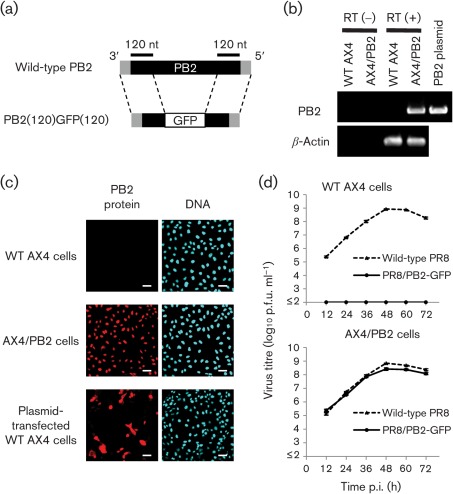
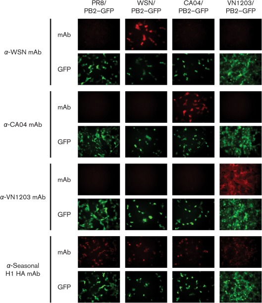
A biologically contained influenza A virus that stably expresses a foreign gene can be effectively traced, used to generate a novel multivalent vaccine and have its replication easily assessed, all while satisfying safety concerns regarding pathogenicity or reversion. This study generated a PB2-knockout (PB2-KO) influenza virus that harboured the GFP reporter gene in the coding region of its PB2 viral RNA (vRNA). Replication of the PB2-KO virus was restricted to a cell line stably expressing the PB2 protein. The GFP gene-encoding PB2 vRNA was stably incorporated into progeny viruses during replication in PB2-expressing cells. The GFP gene was expressed in virus-infected cells with no evidence of recombination between the recombinant PB2 vRNA and the PB2 protein mRNA. Furthermore, other reporter genes and the haemagglutinin and neuraminidase genes of different virus strains were accommodated by the PB2-KO virus. Finally, the PB2-KO virus was used to establish an improved assay to screen neutralizing antibodies against influenza viruses by using reporter gene expression as an indicator of virus infection rather than by observing cytopathic effect. These results indicate that the PB2-KO virus has the potential to be a valuable tool for basic and applied influenza virus research.


Figures

Fig. 1.
Fig. 1.
Characterization of PR8/PB2–GFP virus. (a) Schematic diagram of wild-type PB2 and PB2(120)GFP(120) vRNAs. PB2(120)GFP(120) vRNA possesses the 3′ non-coding region, 120 nt of the coding sequence of PB2 vRNA, the GFP gene and 120 nt of the 3′ and 5′ non-coding regions of PB2 vRNA. The non-coding region and coding regions of PB2 vRNA are represented by shaded and filled bars, respectively. (b) PB2 gene expression in AX4/PB2 cells. RNA was extracted from wild-type AX4 and AX4/PB2 cells. RT-PCR was performed using an oligo(dT) primer followed by cDNA synthesis and PCR with PB2-specific or canine β-actin-specific primers. (c) PB2 protein expression in AX4/PB2 cells. Cells were reacted with anti-PB2 antibody (clone 18/1; left panels) and the nuclei stained with Hoechst 33342 (right panels). Bars, 50 µm. (d) Growth kinetics of PR8/PB2–GFP monitored over 72 h. Wild-type AX4 and AX4/PB2 cells were infected with wild-type PR8 or PR8/PB2–GFP virus at an m.o.i. of 0.001. Supernatants collected at the indicated time points were assayed for infectious virus in plaque assays in AX4/PB2 cells.
Fig. 2.
Fig. 2.
Accommodation of various HA genes in PB2-KO virus and HA expression in PB2-KO virus-infected cells. AX4/PB2 cells were infected with PR8/PB2–GFP, WSN/PB2–GFP, CA04/PB2–GFP or VN1203/PB2–GFP. At 16 h p.i., the cells were stained with mAbs specific for WSN, CA04, VN1203 or seasonal H1 HA protein, respectively. Expression of HA and GFP was examined by fluorescence microscopy.
Fig. 3.
Fig. 3.
Accommodation of various reporter genes in PB2-KO virus. (a) Luciferase activity in PB2-KO virus-infected cells. Wild-type AX4 and AX4/PB2 cells were infected with PR8/PB2–Fluc or PR8/PB2–Rluc at the indicated m.o.i. At 8 h p.i., Fluc and Rluc activities in the cells were measured using a dual-luciferase reporter assay system. (b) GFP intensity in PB2-KO virus-infected cells. AX4/PB2 cells were infected with PR8/PB2–GFP at the indicated m.o.i. At 8 h p.i., GFP intensity was measured using a microplate reader. In (a) and (b), the results from virus-infected cells were compared with those from uninfected cells (indicated by ‘0’) and P values were calculated using Student’s t-test. *, P<0.05. RFU, Relative fluorescent units.
Fig. 4.
Fig. 4.
PB2-KO virus-based microneutralization assay. (a) Growth kinetics of CA04/PB2–Rluc monitored over 72 h. Wild-type AX4 and AX4/PB2 cells were infected with wild-type CA04 or CA04/PB2–Rluc virus at an m.o.i. of 0.001. Supernatants collected at the indicated time points were assayed for infectious virus by plaque assay in AX4/PB2 cells. (b) AX4/PB2 cells were infected with 100 p.f.u. CA04/PB2–Rluc that had been pre-mixed with serially diluted ferret serum samples in triplicate wells. Rluc activity in cells was measured using a Renilla luciferase assay system at 24 h p.i. The results from virus-infected cells were compared with those from cells that were infected with serum-untreated (−) virus. P values were calculated using Student’s t-test. *, P<0.05. RLU, Relative light units.

References

    1. Davies W. L., Grunert R. R., Haff R. F., McGahen J. W., Neumayer E. M., Paulshock M., Watts J. C., Wood T. R., Hermann E. C., Hoffmann C. E. (1964). Antiviral activity of 1-adamantanamine (amantadine). Science 144, 862–863 10.1126/science.144.3620.862 - DOI - PubMed
    1. DuBridge R. B., Tang P., Hsia H. C., Leong P.-M., Miller J. H., Calos M. P. (1987). Analysis of mutation in human cells by using an Epstein–Barr virus shuttle system. Mol Cell Biol 7, 379–387 - PMC - PubMed
    1. Hatakeyama S., Sakai-Tagawa Y., Kiso M., Goto H., Kawakami C., Mitamura K., Sugaya N., Suzuki Y., Kawaoka Y. (2005). Enhanced expression of an α2,6-linked sialic acid on MDCK cells improves isolation of human influenza viruses and evaluation of their sensitivity to a neuraminidase inhibitor. J Clin Microbiol 43, 4139–4146 10.1128/JCM.43.8.4139-4146.2005 - DOI - PMC - PubMed
    1. Hatta M., Asano Y., Masunaga K., Ito T., Okazaki K., Toyoda T., Kawaoka Y., Ishihama A., Kida H. (2000). Mapping of functional domains on the influenza A virus RNA polymerase PB2 molecule using monoclonal antibodies. Arch Virol 145, 1947–1961 10.1007/s007050070068 - DOI - PubMed
    1. Hayden F. G. (2001). Perspectives on antiviral use during pandemic influenza. Philos Trans R Soc Lond B Biol Sci 356, 1877–1884 10.1098/rstb.2001.1007 - DOI - PMC - PubMed

Publication types

MeSH terms