

Experimental model of tuberculosis in the domestic goat after endobronchial infection with Mycobacterium caprae
- PMID: 21880849
- PMCID: PMC3209027
- DOI: 10.1128/CVI.05323-11
Experimental model of tuberculosis in the domestic goat after endobronchial infection with Mycobacterium caprae
Abstract
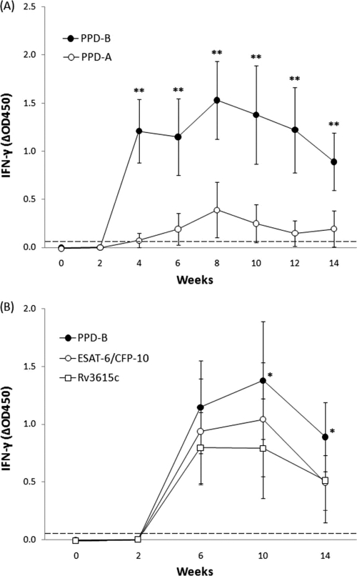
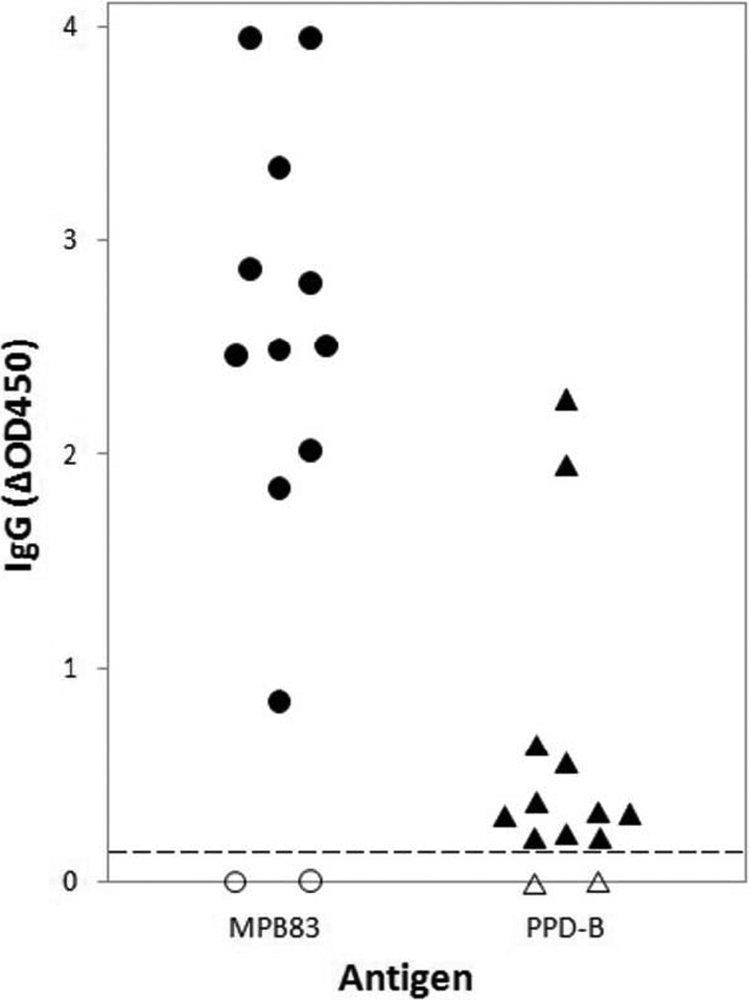
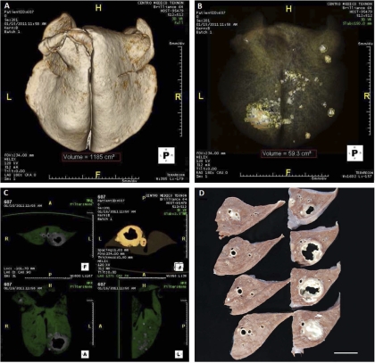
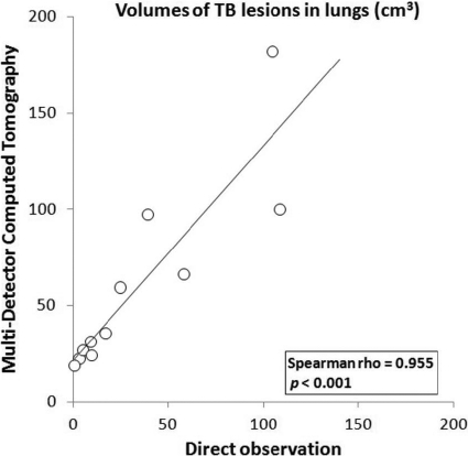
Caprine tuberculosis (TB) has increased in recent years, highlighting the need to address the problem the infection poses in goats. Moreover, goats may represent a cheaper alternative for testing of prototype vaccines in large ruminants and humans. With this aim, a Mycobacterium caprae infection model has been developed in goats. Eleven 6-month-old goats were infected by the endobronchial route with 1.5 × 10(3) CFU, and two other goats were kept as noninfected controls. The animals were monitored for clinical and immunological parameters throughout the experiment. After 14 weeks, the goats were euthanized, and detailed postmortem analysis of lung lesions was performed by multidetector computed tomography (MDCT) and direct observation. The respiratory lymph nodes were also evaluated and cultured for bacteriological analysis. All infected animals were positive in a single intradermal comparative cervical tuberculin (SICCT) test at 12 weeks postinfection (p.i.). Gamma interferon (IFN-γ) antigen-specific responses were detected from 4 weeks p.i. until the end of the experiment. The humoral response to MPB83 was especially strong at 14 weeks p.i. (13 days after SICCT boost). All infected animals presented severe TB lesions in the lungs and associated lymph nodes. M. caprae was recovered from pulmonary lymph nodes in all inoculated goats. MDCT allowed a precise quantitative measure of TB lesions. Lesions in goats induced by M. caprae appeared to be more severe than those induced in cattle by M. bovis over a similar period of time. The present work proposes a reliable new experimental animal model for a better understanding of caprine tuberculosis and future development of vaccine trials in this and other species.
Figures
References
-
- Aranaz A., Cousins D., Mateos A., Dominguez L. 2003. Elevation of Mycobacterium tuberculosis subsp. caprae Aranaz et al. 1999 to species rank as Mycobacterium caprae comb. nov., sp. nov. Int. J. Syst. Evol. Microbiol. 53:1785–1789 - PubMed
-
- Bezos J., et al. 2011. Assessment of in vivo and in vitro tuberculosis diagnostic tests in Mycobacterium caprae naturally infected caprine flocks. Prev. Vet. Med. 100:187–192 - PubMed
-
- Bezos J., et al. 2010. Experimental infection with Mycobacterium caprae in goats and evaluation of immunological status in tuberculosis and paratuberculosis co-infected animals. Vet. Immunol. Immunopathol. 133:269–275 - PubMed
-
- Buddle B. M., Aldwell F. E., Pfeffer A., de Lisle G. W. 1994. Experimental Mycobacterium bovis infection in the brushtail possum (Trichosurus vulpecula): pathology, haematology and lymphocyte stimulation responses. Vet. Microbiol. 38:241–254 - PubMed
-
- Buddle B. M., Wedlock D. N., Denis M. 2006. Progress in the development of tuberculosis vaccines for cattle and wildlife. Vet. Microbiol. 112:191–200 - PubMed
Publication types
MeSH terms
Substances
LinkOut - more resources
Full Text Sources
Medical
