. 2011 Nov;22(21):4029-37.
doi: 10.1091/mbc.E11-03-0260. Epub 2011 Aug 31.

Stimulation of the CLIP-170--dependent capture of membrane organelles by microtubules through fine tuning of microtubule assembly dynamics

Affiliations

Stimulation of the CLIP-170--dependent capture of membrane organelles by microtubules through fine tuning of microtubule assembly dynamics

Alexis J Lomakin et al. Mol Biol Cell. 2011 Nov.

Abstract

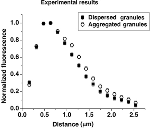
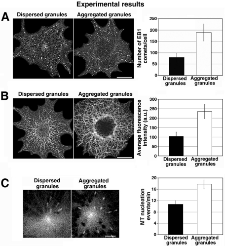
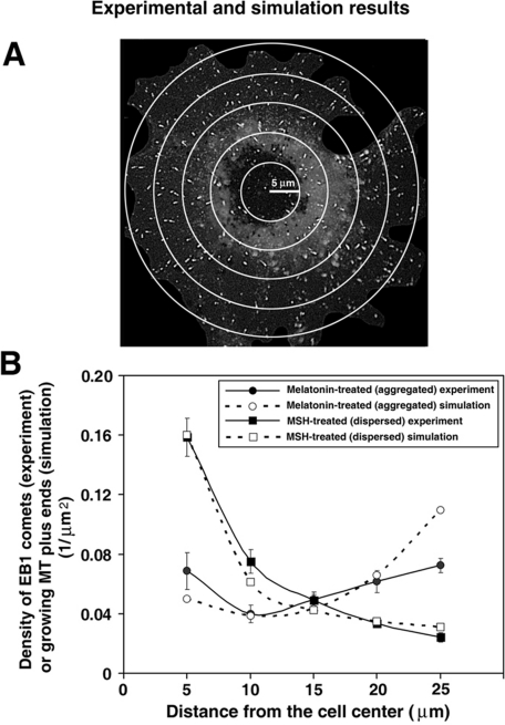
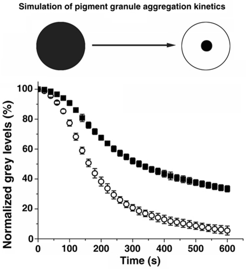
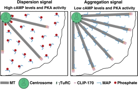
Cytoplasmic microtubules (MTs) continuously grow and shorten at their free plus ends, a behavior that allows them to capture membrane organelles destined for MT minus end-directed transport. In Xenopus melanophores, the capture of pigment granules (melanosomes) involves the +TIP CLIP-170, which is enriched at growing MT plus ends. Here we used Xenopus melanophores to test whether signals that stimulate minus end MT transport also enhance CLIP-170-dependent binding of melanosomes to MT tips. We found that these signals significantly (>twofold) increased the number of growing MT plus ends and their density at the cell periphery, thereby enhancing the likelihood of interaction with dispersed melanosomes. Computational simulations showed that local and global increases in the density of CLIP-170-decorated MT plus ends could reduce the half-time of melanosome aggregation by ~50%. We conclude that pigment granule aggregation signals in melanophores stimulate MT minus end-directed transport by the increasing number of growing MT plus ends decorated with CLIP-170 and redistributing these ends to more efficiently capture melanosomes throughout the cytoplasm.


Figures

FIGURE 1:
FIGURE 1:
Lengths of MT segments decorated with CLIP-170 are similar between cells stimulated to aggregate or disperse pigment granules. Profiles of CLIP-170 fluorescence at MT tips normalized by maximum fluorescence and averaged for cells stimulated to aggregate (open circles) or disperse (filled squares) melanosomes. Stimulation of pigment granule aggregation does not significantly change the length of MT segments decorated by CLIP-170.
FIGURE 2:
FIGURE 2:
Pigment granule aggregation signals increase the total number of growing MT plus ends by stimulating MT nucleation at the centrosome. (A) Immunostaining of melanophores with an antibody against EB1. Left, images of immunostained cells; right, quantification of the average number of MT segments immunostained with EB1 antibody per cell. In melanophores with aggregated melanosomes the number of EB1-labeled segments is significantly higher than in cells with dispersed melanosomes. (B) Immunostaining of methanol-extracted-fixed melanophores with an anti-tubulin antibody. Left, images of immunostained cells; right, quantification of MT fluorescence. MT fluorescence and therefore MT polymer level are substantially higher in cells with aggregated than in those with dispersed melanosomes. (C) Measurement of the frequency of MT nucleation at the centrosome using EB1-GFP. Left, live fluorescence images of EB1-GFP at the centrosome region; right, quantification of the average number of EB1-GFP comets that emerged from the centrosome region per minute. The frequency of EB1-GFP comets that emerged from the centrosome region is higher in cells with aggregated than dispersed melanosomes.
FIGURE 3:
FIGURE 3:
Pigment granule aggregation stimuli increase the density of growing MT plus ends at the cell periphery by changing the parameters of MT dynamic instability. (A) Method for the measurement of density distribution of growing MT plus ends along the cell radius. EB1 comets were counted within each of five regions delineated with concentric circles placed at the same distance from each other over fluorescence images of immunostained cells. (B) EB1 comet counts were normalized for each region area, and data obtained for cells treated to aggregate (circles) or disperse (squares) melanosomes were averaged and used to generate plots of EB1 comet density as a function of distance along the cell radius. Closed symbols represent experimental data. Open symbols show the values determined by computer simulations of MT dynamics based on parameters of MT dynamic instability. Error bars represent SE of the mean. Computational and experimental data agree with each other and show that in melanophores with dispersed melanosomes the density of growing MT plus ends decreases with increasing distance from the cell center, whereas in cells stimulated to aggregate pigment granules, growing MT plus end density is approximately similar at the cell center and the cell periphery.
FIGURE 4:
FIGURE 4:
Changes in MT dynamic instability and nucleation induced by pigment granule aggregation stimuli significantly increase the rate of pigment granule aggregation. Top, cartoon illustrating the principle of the method used for computing the kinetics of pigment granule aggregation. In the dispersed state, a homogeneous pigment granule distribution makes the cytoplasm dark. In this state, gray level (the measure of darkness of the cytoplasm) is designated to be 100%. During aggregation, the cytoplasm becomes increasingly transparent, and gray level drops to a fraction of the initial value. Bottom, comparison of pigment granule aggregation kinetics. Open circles represent the gray level kinetics in a virtual melanophore with the granule movement and MT parameters associated with aggregation; black squares represent the gray level kinetics in a virtual melanophore with the granule movement parameters associated with aggregation and the MT parameters associated with dispersion. Data are expressed as the gray level percentage change with time. The error bars reflect the mean square displacement over at least 10 simulations per parameter set. Microtubule parameters associated with aggregation result in a significantly faster gray level decrease.
FIGURE 5:
FIGURE 5:
Model for MT regulation in melanophores. Left, in cells stimulated to disperse pigment granules, cAMP levels and the activity of PKA are high, which leads to phosphorylation and inactivation of centrosomal proteins involved in MT nucleation, such as γTuRC, and cytoplasmic microtubule-associated proteins responsible for MT polymerization, which reduces the number of growing MT plus ends and decreases their density at the cell periphery. Right, pigment granule aggregation signals decrease cAMP levels and PKA activity, which relieves the PKA-dependent inhibition of MT nucleation and growth. As a result, more MTs nucleate at the centrosome and persistently grow to the cell periphery, thus increasing the total number and local density of MT ends at the cell periphery. Increases in the local density of growing MT plus ends at the cell periphery and their total number throughout the cytoplasm enhance pigment granule aggregation.

References

    1. Akhmanova A, Steinmetz MO. Tracking the ends: a dynamic protein network controls the fate of microtubule tips. Nat Rev Mol Cell Biol. 2008;9:309–322. - PubMed
    1. Andersen SS. Molecular characteristics of the centrosome. Int Rev Cytol. 1999;187:51–109. - PubMed
    1. Aspengren S, Hedberg D, Skold HN, Wallin M. New insights into melanosome transport in vertebrate pigment cells. Int Rev Cell Mol Biol. 2009;272:245–302. - PubMed
    1. Belmont LD, Mitchison TJ. Identification of a protein that interacts with tubulin dimers and increases the catastrophe rate of microtubules. Cell. 1996;84:623–631. - PubMed
    1. Bieling P, Kandels-Lewis S, Telley IA, van Dijk J, Janke C, Surrey T. CLIP-170 tracks growing microtubule ends by dynamically recognizing composite EB1/tubulin-binding sites. J Cell Biol. 2008;183:1223–1233. - PMC - PubMed

Publication types

MeSH terms

LinkOut - more resources