. 2011 Aug 31;31(35):12426-36.
doi: 10.1523/JNEUROSCI.0685-11.2011.

Essential role for vav Guanine nucleotide exchange factors in brain-derived neurotrophic factor-induced dendritic spine growth and synapse plasticity

Affiliations

Essential role for vav Guanine nucleotide exchange factors in brain-derived neurotrophic factor-induced dendritic spine growth and synapse plasticity

Carly F Hale et al. J Neurosci. .

Abstract

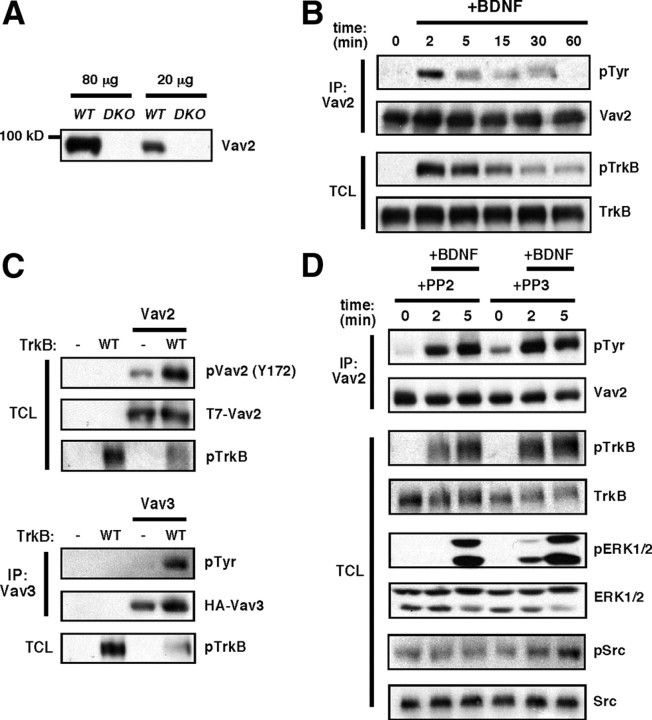
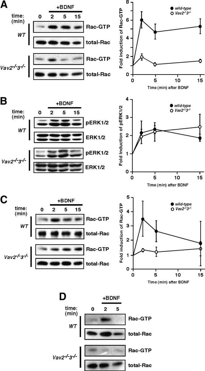
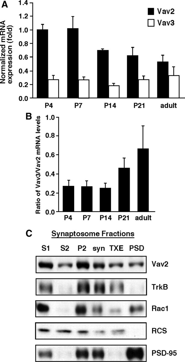
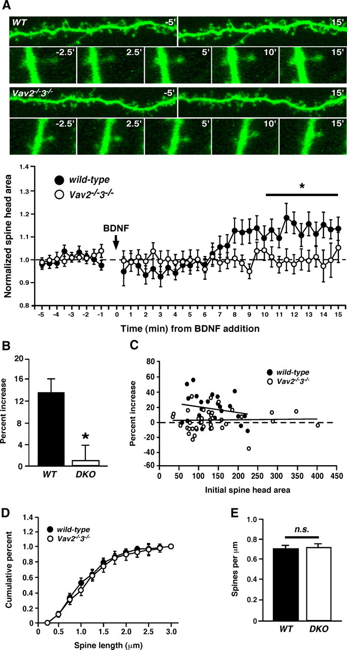
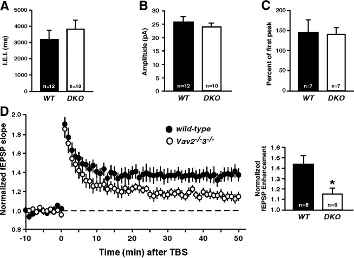
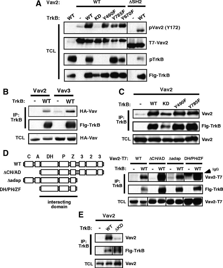
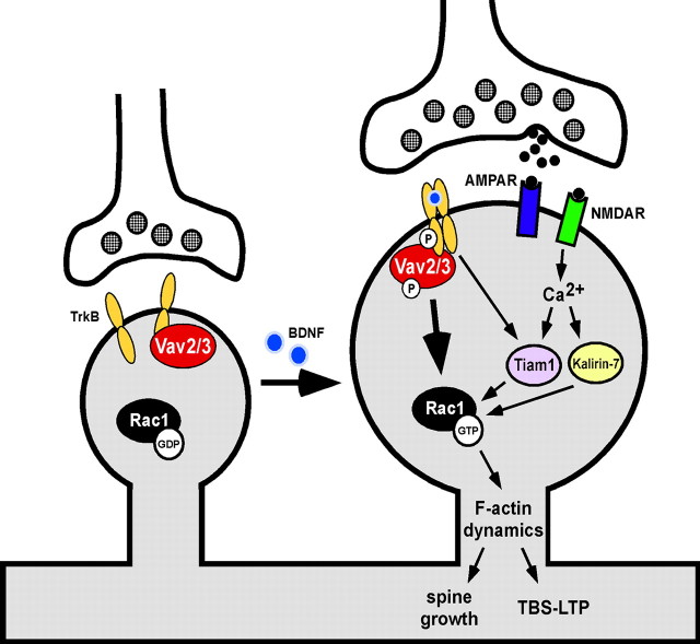
Brain-derived neurotrophic factor (BDNF) and its cognate receptor, TrkB, regulate a wide range of cellular processes, including dendritic spine formation and functional synapse plasticity. However, the signaling mechanisms that link BDNF-activated TrkB to F-actin remodeling enzymes and dendritic spine morphological plasticity remain poorly understood. We report here that BDNF/TrkB signaling in neurons activates the Vav family of Rac/RhoA guanine nucleotide exchange factors through a novel TrkB-dependent mechanism. We find that Vav is required for BDNF-stimulated Rac-GTP production in cortical and hippocampal neurons. Vav is partially enriched at excitatory synapses in the postnatal hippocampus but does not appear to be required for normal dendritic spine density. Rather, we observe significant reductions in both BDNF-induced, rapid, dendritic spine head growth and in CA3-CA1 theta burst-stimulated long-term potentiation in Vav-deficient mouse hippocampal slices, suggesting that Vav-dependent regulation of dendritic spine morphological plasticity facilitates normal functional synapse plasticity.


Figures

Figure 1.
Figure 1.
BDNF stimulates the transient activation of Vav2 in neurons. A, Vav2 antibodies are highly specific. Western blot of whole hippocampi from P15 WT or Vav2−/−3−/− (DKO) mice with anti-Vav2 antibody (Cowan et al., 2005). B, E18 rat cortical neurons were cultured for 6 d and then stimulated with BDNF (100 ng/ml) over a time course. Vav2 was immunoprecipitated and blotted with antiphosphotyrosine antibody (4G10). Total cell lysates (TCLs) were blotted with anti-phospho-TrkB (Y490) and anti-TrkB antibodies. The results are a representative finding of five independent experiments. C, Coexpression of Vav2 or Vav3 and TrkB increases phospho-Vav in HEK293T cells. HEK293T cells were transfected with T7-tagged Vav2 or HA-tagged Vav3 and Flag-tagged TrkB. To examine Vav2 Y172 phosphorylation, whole-cell lysates were blotted with anti-phospho-Vav2 (Y172) antibody. To examine Vav3 phosphorylation, total cell lysates were immunoprecipitated with anti-HA antibody, then immunoblotted with antiphosphotyrosine (4G10) and anti-HA (Vav) antibodies. Total cell lysates were blotted with anti-pTrkB (Y490) antibody. The results are a representative finding of three independent experiments. D, BDNF stimulates activation of Vav2 independently of Src kinase activity. E18 cortical neurons were cultured for 6 d and then incubated with DMSO, PP2 (5 μm), or PP3 (5 μm) for 30 min before stimulation with BDNF (100 ng/ml) over a time course. Vav2 was immunoprecipitated and blotted with antiphosphotyrosine antibody (4G10). Total cell lysates were blotted with anti-phospho-TrkB (Y490), anti-TrkB, anti-phospho-ERK1/2 (T202/Y204), anti-ERK1/2, anti-phospho-Src (Y418), and anti-Src antibodies. The results are a representative finding of two independent experiments.
Figure 2.
Figure 2.
BDNF stimulates Rac-GTP formation through a Vav-dependent mechanism. A, Hippocampal organotypic slices (400 μm) were cultured from wild-type or Vav2−/−3−/− mice at P6. At 9 DIV, slices were stimulated with BDNF (250 ng/ml) over a time course. Rac1-GTP was precipitated using GST-PBD fusion protein and glutathione Sepharose. Total cell lysates were blotted with anti-Rac1 antibody. Rac1-GTP was normalized to total Rac1 levels and expressed as a fold change compared with the 0 min time point (p < 0.001, genotype by two-way ANOVA). Wild-type and Vav2−/−3−/− data are from four and five independent experiments, respectively. B, Erk1/2 pathway is activated normally in Vav2−/−3−/− hippocampi (as in A) in response to BDNF stimulation. Total cell lysates were blotted with anti-phospho-ERK1/2 (T202/Y204) and anti-ERK1/2 antibodies. Wild-type and Vav2−/−3−/− data are from three and four independent experiments, respectively. C, Rac1-GTP stimulation by BDNF was performed as in A, except that cultured primary cortical neurons (E16.5 + 6DIV) from either wild-type or Vav2−/−3−/− mice were used and treatment was administered with 100 ng/ml BDNF for the indicated times. Wild-type and Vav2−/−3−/− data are from four independent experiments. D, Acute hippocampal slices (300 μm) from adult (3 month) wild-type or Vav2−/−3−/− mice were stimulated with BDNF (100 ng/ml) over a time course. Rac1-GTP was precipitated using GST-PBD fusion protein and glutathione Sepharose. Wild-type and Vav2−/−3−/− results are representative findings from three independent experiments.
Figure 3.
Figure 3.
Vav GEFs colocalize with excitatory synaptic proteins in the hippocampus. A, Relative hippocampal expression of Vav2 and Vav3 mRNA at indicated time points was determined using real-time qPCR. Results were normalized to cyclophilin mRNA levels. For A and B, data are from three independent experiments, each of which was conducted in triplicate. B, Ratio of Vav3/Vav2 mRNA expression at indicated time points. C, Synaptosomes were prepared from whole hippocampi of P15 mice. Vav2 localized to the membrane (P2) and synaptosome fractions. S1, Total hippocampal homogenate with nuclei removed; S2, cytosol; P2, crude membrane fraction; syn, synaptosomes collected following Ficoll gradient fractionation; TXE, 1% Triton X-100-soluble fraction of syn; PSD, pellet after 0.5% Triton X-100 extraction. Synaptosome fractions were blotted with anti-Vav2, anti-TrkB, anti-Rac, anti-RCS (regulator of calmodulin signaling) (Pulipparacharuvil et al., 2008), and anti-PSD-95 antibodies. The results are a representative finding of two independent experiments.
Figure 4.
Figure 4.
Vav GEFs are required for BDNF-induced dendritic spine head growth in hippocampal CA1 neurons. A, Top, Hippocampal organotypic slices (400 μm) were cultured from wild-type or Vav2−/−3−/− mice at P6. At 9 DIV, slices were stimulated with BDNF (250 ng/ml) over a time course and imaged using 2 photon microscopy. Representative photomicrographs showing dendritic segments and dendritic spine heads from wild-type or Vav2−/−3−/− mice at indicated times from BDNF stimulation. Data plot: normalized spine head areas from wild-type and Vav2−/−3−/− mice at times before and after BDNF addition to slice. *p < 0.05, two-way ANOVA; genotype and time, n = 6 (wild-type), n = 8 (Vav2−/−3−/−). For all experiments (A–E), n = 6 (wild-type) and n = 8 (Vav2−/−3−/−). Each n represents an average of five randomly chosen dendritic spines from one dendritic length. One dendritic length per slice was imaged and averaged to give an average spine behavior per dendrite. B, Percentage increase of mean spine head size at 10–15 min post-BDNF compared with the first 5 min after BDNF application (*p < 0.05, Student's t test). C, Plot of the percentage increase in spine head size versus initial spine head area, demonstrating that a similar range of spine heads was analyzed and that the failure of Vav KO spine head growth with BDNF treatment is not due to occlusion. D, Plot of cumulative dendritic spine length of wild-type and Vav2−/−3−/− neurons from pre-BDNF treated spines in A. E, Dendritic spine densities of wild-type and Vav2−/−3−/− neurons, cultured and imaged in A.
Figure 5.
Figure 5.
Vav GEFs mediate synaptic plasticity in the hippocampus. A, Average IEI per neuron from wild-type or Vav2−/−3−/− CA1 pyramidal neurons (n.s., Student's t test). B, Average mEPSC amplitude per neuron from same recordings as in B (n.s., Student's t test). C, Paired-pulse stimulation (10 Hz). Average paired-pulse ratio measured as the percentage of the second pulse relative to the first (n.s., Student's t test). D, Left, fEPSPs were recorded from CA1 of P15 wild-type or Vav2−/−3−/− acute hippocampal slices (300 μm) in response to two theta burst stimulations of Schaffer collaterals. Right, The fEPSP slopes were normalized for each genotype. Normalized fEPSP enhancement at 40–50 min is shown (*p < 0.05, Student's t test).
Figure 6.
Figure 6.
Kinase-active TrkB activates Vav2 independently of the ERK/PI3K- and PLCγ-interacting sites. A, Coexpression of TrkB and Vav2 increases phospho-Vav2 (Y172) in HEK293T cells. HEK293T cells were transfected with T7-tagged wild-type or ΔSH2 and Flag-tagged wild-type or mutant TrkB. Total cell lysates were blotted with anti-phospho-Vav2 (Y172), anti-T7 (Vav2), anti-phospho-TrkB (Y490), and anti-TrkB antibodies. A–E, Results are representative findings from three independent experiments. B, TrkB binds Vav2 and Vav3 in HEK293T cells. HEK293T cells were transfected with HA-tagged Vav2 or Vav3 and Flag-tagged TrkB. Total cell lysates were immunoprecipitated with anti-Flag antibody, then immunoblotted with anti-HA (Vav) and anti-Flag (TrkB) antibodies. Whole-cell lysates were blotted with anti-HA (Vav) antibody. C, TrkB binds Vav2 independently of TrkB kinase activity. T7-tagged Vav2 was coexpressed with Flag-tagged TrkB (wild-type), TrkB-KD (K571N), TrkB-Y490F, or TrkB-Y785F. Total cell lysates (TCLs) were immunoprecipitated with anti-Flag antibody, then immunoblotted with anti-Vav2 and anti-Flag (TrkB) antibodies. Whole-cell lysates were blotted with anti-Vav2 antibody. D, Deletion analysis of Vav2 identifies the DH/PH/ZF region as sufficient to mediate coassociation with TrkB. Domain composition of full-length and Vav2 deletion mutants that were generated. C, Calponin homology domain; A, acidic domain; DH, Dbl homology domain; P, pleckstrin homology domain; Z, zinc finger domain; P, proline-rich domain; 3, Src homology type 3 domain (SH3); 2, Src homology type 2 domain (SH2). HEK293T cells were transfected with Flag-tagged TrkB (wild-type) and T7-tagged Vav2 (wild-type), Vav2-ΛCH/AD, Vav2-Λadaptor, or Vav2-DH/PH/ZF. Total cell lysates were immunoprecipitated with anti-Flag antibody and immunoblotted with anti-T7 (Vav2) and anti-Flag (TrkB) antibodies. Whole-cell lysate was blotted with anti-T7 (Vav2) antibody. E, TrkB's kinase domain is required for its interaction with Vav2. T7-tagged Vav2 was coexpressed with wild-type or Δ kinase domain (ΔKD)-TrkB. Total cell lysates were immunoprecipitated with anti-Flag antibody and immunoblotted with anti-Vav2 and anti-Flag (TrkB) antibodies. Whole-cell lysate was blotted with anti-Vav2 antibody.
Figure 7.
Figure 7.
Model for the role of Vav GEFs in BDNF/TrkB-induced dendritic spine growth and functional synapse plasticity.

Similar articles

Cited by

References

    1. Abe K, Rossman KL, Liu B, Ritola KD, Chiang D, Campbell SL, Burridge K, Der CJ. Vav2 is an activator of Cdc42, Rac1, and RhoA. J Biol Chem. 2000;275:10141–10149. - PubMed
    1. Aghazadeh B, Lowry WE, Huang XY, Rosen MK. Structural basis for relief of autoinhibition of the Dbl homology domain of proto-oncogene Vav by tyrosine phosphorylation. Cell. 2000;102:625–633. - PubMed
    1. Bramham CR. Local protein synthesis, actin dynamics, and LTP consolidation. Curr Opin Neurobiol. 2008;18:524–531. - PubMed
    1. Bustelo XR. Regulatory and signaling properties of the Vav family. Mol Cell Biol. 2000;20:1461–1477. - PMC - PubMed
    1. Bustelo XR. Vav proteins, adaptors and cell signaling. Oncogene. 2001;20:6372–6381. - PubMed

Publication types

MeSH terms

Substances