

Inhibitory dendrite dynamics as a general feature of the adult cortical microcircuit
- PMID: 21880904
- PMCID: PMC3180878
- DOI: 10.1523/JNEUROSCI.0420-11.2011
Inhibitory dendrite dynamics as a general feature of the adult cortical microcircuit
Abstract
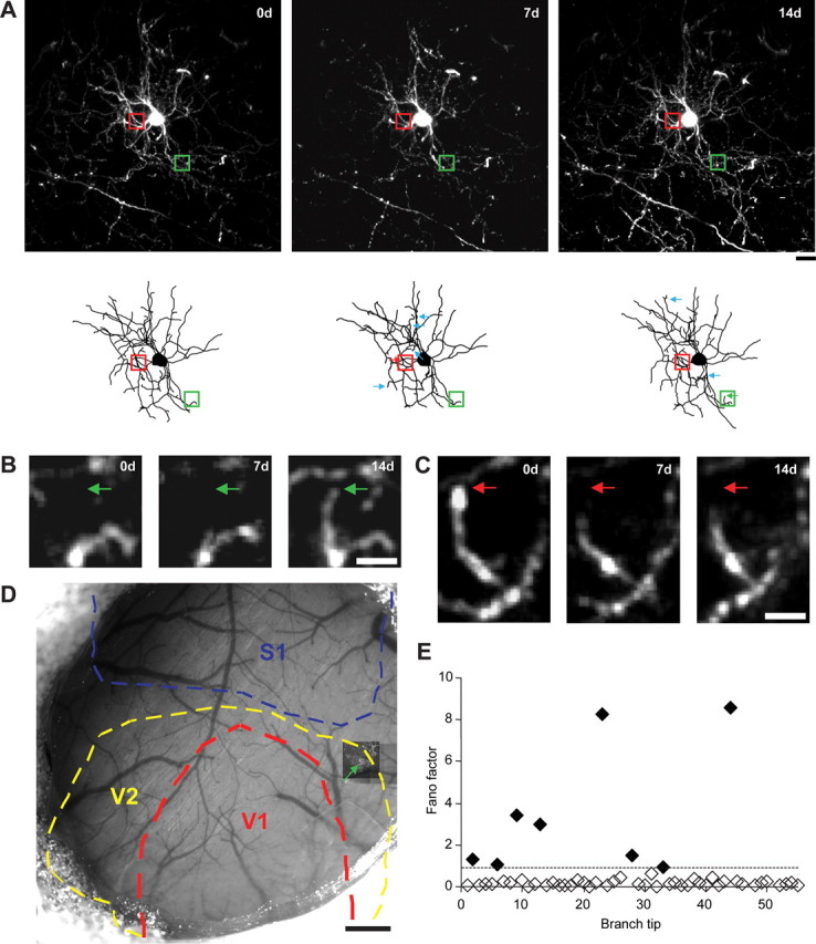
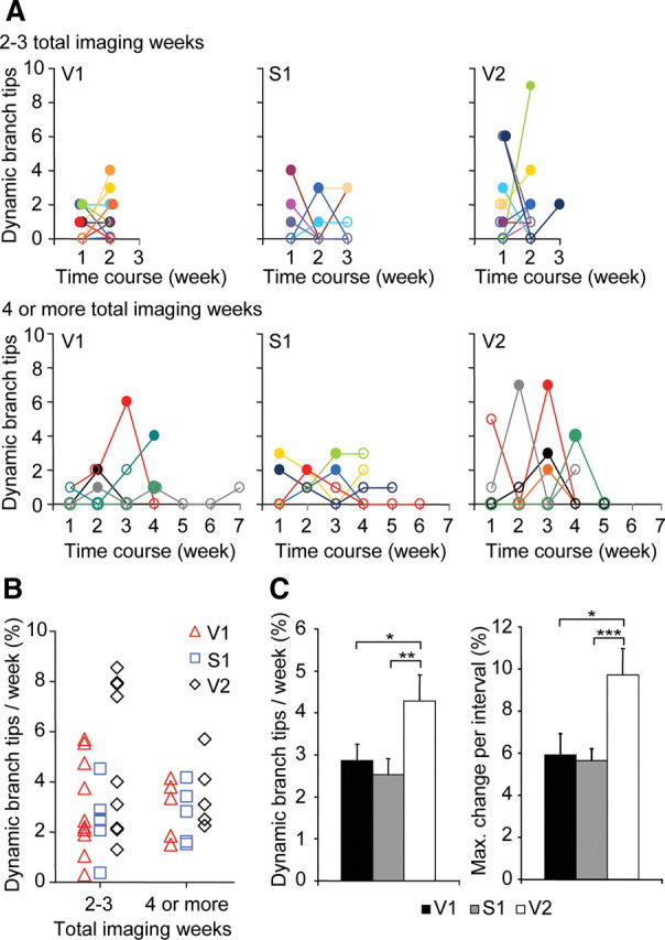
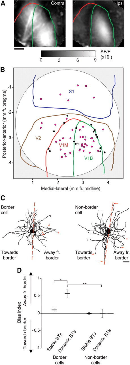
The mammalian neocortex is functionally subdivided into architectonically distinct regions that process various types of information based on their source of afferent input. Yet, the modularity of neocortical organization in terms of cell type and intrinsic circuitry allows afferent drive to continuously reassign cortical map space. New aspects of cortical map plasticity include dynamic turnover of dendritic spines on pyramidal neurons and remodeling of interneuron dendritic arbors. While spine remodeling occurs in multiple cortical regions, it is not yet known whether interneuron dendrite remodeling is common across primary sensory and higher-level cortices. It is also unknown whether, like pyramidal dendrites, inhibitory dendrites respect functional domain boundaries. Given the importance of the inhibitory circuitry to adult cortical plasticity and the reorganization of cortical maps, we sought to address these questions by using two-photon microscopy to monitor interneuron dendritic arbors of thy1-GFP-S transgenic mice expressing GFP in neurons sparsely distributed across the superficial layers of the neocortex. We find that interneuron dendritic branch tip remodeling is a general feature of the adult cortical microcircuit, and that remodeling rates are similar across primary sensory regions of different modalities, but may differ in magnitude between primary sensory versus higher cortical areas. We also show that branch tip remodeling occurs in bursts and respects functional domain boundaries.
Figures
Similar articles
-
Interneuron Simplification and Loss of Structural Plasticity As Markers of Aging-Related Functional Decline.J Neurosci. 2018 Sep 26;38(39):8421-8432. doi: 10.1523/JNEUROSCI.0808-18.2018. Epub 2018 Aug 14. J Neurosci. 2018. PMID: 30108129 Free PMC article.
-
Structural basis for the role of inhibition in facilitating adult brain plasticity.Nat Neurosci. 2011 May;14(5):587-94. doi: 10.1038/nn.2799. Epub 2011 Apr 10. Nat Neurosci. 2011. PMID: 21478885 Free PMC article.
-
Novel interneuronal network in the mouse posterior piriform cortex.J Comp Neurol. 2006 Dec 20;499(6):1000-15. doi: 10.1002/cne.21166. J Comp Neurol. 2006. PMID: 17072835
-
Highly specific structural plasticity of inhibitory circuits in the adult neocortex.Neuroscientist. 2013 Aug;19(4):384-93. doi: 10.1177/1073858413479824. Epub 2013 Mar 8. Neuroscientist. 2013. PMID: 23474602 Free PMC article. Review.
-
Sensory experience and cortical rewiring.Neuroscientist. 2010 Apr;16(2):186-98. doi: 10.1177/1073858409343961. Epub 2009 Oct 1. Neuroscientist. 2010. PMID: 19801372 Review.
Cited by
-
Experience-Dependent Development of Dendritic Arbors in Mouse Visual Cortex.J Neurosci. 2020 Aug 19;40(34):6536-6556. doi: 10.1523/JNEUROSCI.2910-19.2020. Epub 2020 Jul 15. J Neurosci. 2020. PMID: 32669356 Free PMC article.
-
NMDA Receptors Regulate the Structural Plasticity of Spines and Axonal Boutons in Hippocampal Interneurons.Front Cell Neurosci. 2017 Jun 12;11:166. doi: 10.3389/fncel.2017.00166. eCollection 2017. Front Cell Neurosci. 2017. PMID: 28659763 Free PMC article.
-
Interneuron Simplification and Loss of Structural Plasticity As Markers of Aging-Related Functional Decline.J Neurosci. 2018 Sep 26;38(39):8421-8432. doi: 10.1523/JNEUROSCI.0808-18.2018. Epub 2018 Aug 14. J Neurosci. 2018. PMID: 30108129 Free PMC article.
-
Activity-dependent NPAS4 expression and the regulation of gene programs underlying plasticity in the central nervous system.Neural Plast. 2013;2013:683909. doi: 10.1155/2013/683909. Epub 2013 Aug 18. Neural Plast. 2013. PMID: 24024041 Free PMC article. Review.
-
Structural plasticity of interneurons in the adult brain: role of PSA-NCAM and implications for psychiatric disorders.Neurochem Res. 2013 Jun;38(6):1122-33. doi: 10.1007/s11064-013-0977-4. Epub 2013 Jan 26. Neurochem Res. 2013. PMID: 23354722 Review.
References
-
- Buonomano DV, Merzenich MM. Cortical plasticity: from synapses to maps. Annu Rev Neurosci. 1998;21:149–186. - PubMed
Publication types
MeSH terms
Substances
Grants and funding
LinkOut - more resources
Full Text Sources
Miscellaneous