. 2011 Aug 31;31(35):12523-32.
doi: 10.1523/JNEUROSCI.1527-11.2011.

Compartmentalization of the GABAB receptor signaling complex is required for presynaptic inhibition at hippocampal synapses

Affiliations

Compartmentalization of the GABAB receptor signaling complex is required for presynaptic inhibition at hippocampal synapses

Tal Laviv et al. J Neurosci. .

Erratum in

  • J Neurosci. 2012 Feb 15;32(7):2578

Abstract

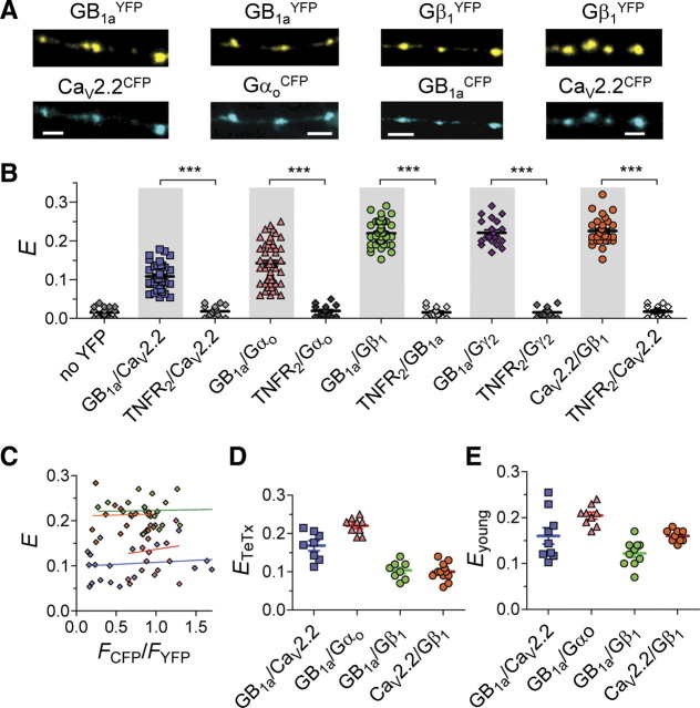
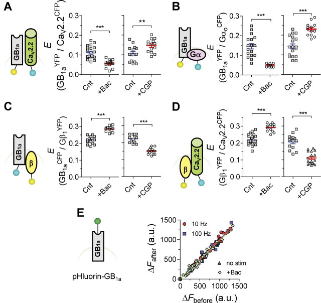
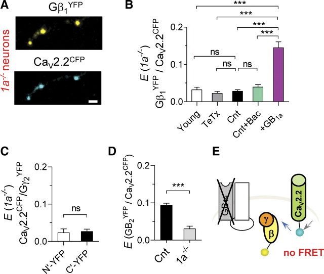
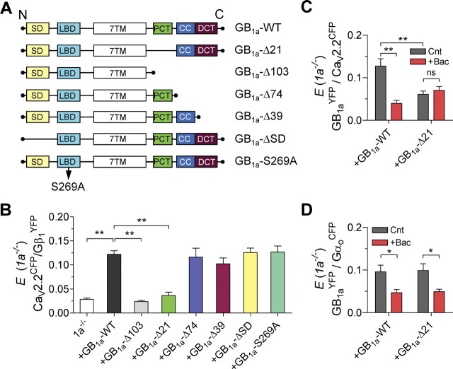
Presynaptic inhibition via G-protein-coupled receptors (GPCRs) and voltage-gated Ca(2+) channels constitutes a widespread regulatory mechanism of synaptic strength. Yet, the mechanism of intermolecular coupling underlying GPCR-mediated signaling at central synapses remains unresolved. Using FRET spectroscopy, we provide evidence for formation of spatially restricted (<100 Å) complexes between GABA(B) receptors composed of GB(1a)/GB(2) subunits, Gα(o)β(1)γ(2) G-protein heterotrimer, and Ca(V)2.2 channels in hippocampal boutons. GABA release was not required for the assembly but for structural reorganization of the precoupled complex. Unexpectedly, GB(1a) deletion disrupted intermolecular associations within the complex. The GB(1a) proximal C-terminal domain was essential for association of the receptor, Ca(V)2.2 and Gβγ, but was dispensable for agonist-induced receptor activation and cAMP inhibition. Functionally, boutons lacking this complex-formation domain displayed impaired presynaptic inhibition of Ca(2+) transients and synaptic vesicle release. Thus, compartmentalization of the GABA(B1a) receptor, Gβγ, and Ca(V)2.2 channel in a signaling complex is required for presynaptic inhibition at hippocampal synapses.


Figures

Figure 1.
Figure 1.
GB1aRs, Gαoβ1γ2 G-protein subunits, and CaV2.2 channels are precoupled at single hippocampal boutons. A, Representative confocal images of pyramidal neuron axons in hippocampal cultures that were cotransfected with GB1aYFP/CaV2.2CFP, GB1aYFP/GαoCFP, GB1aCFP/Gβ1YFP and CaV2.2CFP/Gβ1YFP. Scale bars, 2 μm. B, FRET was detected between GB1aYFP/CaV2.2CFP (n = 33, N = 7), GB1aYFP/Gαo*94CFP (n = 53), GB1aCFP/Gβ1YFP (n = 45, N = 10), GB1aCFP/Gγ2YFP (n = 21, N = 4), CaV2.2CFP/Gβ1YFP (n = 31, N = 6) proteins under miniature synaptic activity at single hippocampal boutons. To verify FRET specificity, E was measured between the CFP-tagged proteins of interest and nonrelated TNFR2YFP. Error bars indicate SEM. ***p < 0.001. C, FRET efficiency is plotted for individual presynaptic boutons as function of CFP/YFP intensity ratio (FCFP/FYFP). No correlation was found: Spearman r is 0.14, 0.11, −0.08, and 0.05 for GB1aYFP/CaV2.2CFP, GB1aYFP/Gαo*94CFP, GB1aCFP/Gβ1YFP, and CaV2.2CFP/Gβ1YFP, respectively (p > 0.5). D, FRET was detected between GB1aYFP/CaV2.2CFP (n = 8, N = 3), GB1aYFP/GαoCFP (n = 9, N = 3), GB1aCFP/Gβ1YFP (n = 8, N = 3), and CaV2.2CFP/Gβ1YFP (n = 12, N = 4) proteins in nonreleasing TeTx-pretreated hippocampal boutons. E, FRET was detected between GB1aYFP/CaV2.2CFP (n = 9, N = 3), GB1aYFP/GαoCFP (n = 9, N = 3), GB1aCFP/Gβ1YFP (n = 10, N = 3), and CaV2.2CFP/Gβ1YFP (n = 9, N = 3) proteins in nonreleasing immature (4–5 DIV) hippocampal neurons.
Figure 2.
Figure 2.
Agonist-induced structural rearrangements in the GB1aR/G-protein/CaV2.2 channel complex. A, E between GB1aYFP/CaV2.2CFP was reduced by baclofen (10 μm, left, n = 13–29, N = 4–6, ***p < 0.0001), but was increased by CGP54626 (1 μm, right, n = 14, N = 4, **p < 0.01). B, E between GB1aYFP/GαoCFP was reduced by baclofen (10 μm, left, n = 12–17, N = 4, ***p < 0.0001), but was increased by GABABR antagonist CGP35348 (1 μm, right, n = 13–18, N = 4, ***p < 0.0001). C, E between GB1aCFP/Gβ1YFP was increased by baclofen (10 μm, left, n = 12–15, N = 4, ***p < 0.0001), but was decreased by GABABR antagonist CGP54626 (1 μm, right, n = 15, N = 4, ***p < 0.0001). D, E between Gβ1YFP/CaV2.2CFP was increased by baclofen (10 μm, left, n = 14–27, N = 4–6, ***p < 0.0001), but was decreased by CGP54626 (1 μm, right, n = 14–17, N = 4–5, ***p < 0.0001). Error bars indicate SEM. E, Fluorescence intensity of pHluorin tagged to GB1a does not change under miniature activity, by application of 10 μm baclofen and as function of stimulation frequency (10 and 100 Hz). Slope of linear fit is 1.04, 0.98, 1.01, and 0.99 for miniature activity, baclofen application, 10 and 100 Hz, respectively.
Figure 3.
Figure 3.
Disruption of FRET between the receptor, CaV2.2 channel, and Gβγ at hippocampal boutons of 1a−/− neurons. A, Confocal images of axonal part of 1a−/− hippocampal pyramidal neuron that was cotransfected with CaV2.2CFP and Gβ1YFP. Scale bar, 2 μm. B, Lack of specific FRET between CaV2.2CFP and Gβ1N′-YFP in 1a−/− boutons: under miniature synaptic activity (Cnt, n = 31, N = 6), in the presence of 10 μm baclofen (n = 16, N = 4), and in TeTx-treated (n = 14, N = 3) and young (n = 14, N = 4) neurons. Transfection of 1a−/− neurons with GB1a resulted in rescue of CaV2.2CFP/Gβ1N′-YFP FRET (n = 17, N = 4, ***p < 0.0001). C, Lack of specific FRET between CaV2.2CFP and either Gγ2N′-YFP (n = 10, N = 3) or Gγ2C′-YFP (n = 9, N = 3). D, Disruption of specific FRET between CaV2.2CFP and GB2YFP protein in 1a−/− neurons (n = 8, N = 3, ***p < 0.0001). E, Diagram illustrating disruption of CaV2.2CFP/Gβ1YFP FRET in boutons of 1a−/− neurons. One-way ANOVA analysis with post hoc Bonferroni's multiple comparison tests (B) and paired t test (C, D) indicated significance. Error bars indicate SEM.
Figure 4.
Figure 4.
Proximal C-terminal domain of the GB1a protein is essential for Gβγ/CaV2.2 channel association. A, Schematics show GB1a constructs used to examine the domain responsible for Gβγ/CaV2.2 association. SD, Two sushi domains; LBD, ligand-binding domain; 7TM, seven-transmembrane domain; PCT, proximal C-terminal domain; CC, coiled–coiled domain; DCT, distal C-terminal domain. B, Mean FRET for the indicated transfection conditions in 1a−/− neurons: GB1a-WT (n = 66, N = 11), GB1a-Δ103 (n = 44, N = 8, **p < 0.01), GB1a-Δ21 (n = 24, N = 6, **p < 0.0001), GB1a-Δ74 (n = 14, N = 4, p > 0.05), GB1a-Δ39 (n = 17, N = 4, p > 0.05), GB1a-ΔSD (n = 15, N = 4, p > 0.05), and GB1a-S269A (n = 14, N = 4, p > 0.05). One-way ANOVA analysis with post hoc Dunnett's multiple-comparison tests relative to 1a−/− boutons transfected with GB1a-WT indicated significance. C, Effect of 10 μm baclofen on the GB1aYFP/CaV2.2CFP FRET in 1a−/− boutons transfected with GB1a-WT (n = 7–20, N = 3–5, **p < 0.01) or GB1a-Δ21 (n = 23–24, N = 5–6, p > 0.05). D, Effect of 10 μm baclofen on the GB1aYFP/GαoCFP FRET in 1a−/− boutons transfected with GB1a-WT (n = 12–15, N = 3–4, *p < 0.05) or GB1a-Δ21 (n = 14–16, N = 3–5, *p < 0.05, paired t test). Error bars indicate SEM.
Figure 5.
Figure 5.
The GB1a proximal C-terminal domain does not affect baclofen-induced GB1a/GB2 receptor activation and cAMP inhibition. A, Dose–response curves of baclofen on GB1aCFP/GB2YFP FRET efficiency for GB1a-WT (n = 10–21, N = 5, ED50 = 0.82 ± 0.003 μm) and GB1a-Δ21 (n = 11–21, N = 3–5, ED50 = 0.58 ± 0.013 μm) proteins. E at 100 μm baclofen was set as 100%. B, Effect of 10 μm baclofen on CFP–Epac–YFP FRET efficiency (EEpac), reporting cAMP level, in 1a−/− boutons and in 1a−/− boutons expressing GB1a-WT or GB1a-Δ21 proteins. Baclofen increased EEpac in GB1a-WT-expressing (n = 30–32, N = 6, *p < 0.05) and GB1a-Δ21-expressing (n = 27–35, N = 5–6, *p < 0.05) boutons, but did not affect EEpac in 1a−/− boutons (n = 25–26, N = 5, p > 0.05) One-way ANOVA with post hoc Bonferroni's multiple-comparison tests indicated significance. Error bars indicate SEM.
Figure 6.
Figure 6.
The role of the GB1a proximal C-terminal domain on baclofen-induced inhibition of Ca2+ transients. A, Baclofen did not affect spike-dependent presynaptic Ca2+ transients (ΔF/F) evoked by 0.2 Hz stimulation in 1a−/− boutons and in GB1a-Δ21-expressing boutons, but reduced it in GB1a-WT-expressing boutons. Ca2+ transients were quantified as before (black) and after (gray) baclofen application (average of 10 traces). B, Average data on baclofen-induced modification in Ca2+ transients in 1a−/− (n = 31, N = 6, p > 0.05), GB1a-WT-expressing (n = 17, N = 4, ***p < 0.001, compared with 1a−/−), and GB1a-Δ21-expressing (n = 19, N = 5, p > 0.05, compared with 1a−/−) boutons. One-way ANOVA with post hoc Bonferroni's multiple-comparison tests indicated significance. Error bars indicate SEM.
Figure 7.
Figure 7.
The GB1a proximal C-terminal domain is required for baclofen-induced inhibition of synaptic vesicle release. A–E, Representative FM destaining curves before and after application of 10 μm baclofen in WT cultures (n = 84, A), WT cultures transfected with MyrPhd and GFP (n = 112, B), 1a−/− cultures (n = 71, C), 1a−/− cultures transfected with GB1a-WT and GFP (n = 69, D), and 1a−/− cultures transfected with GB1a-Δ21 and GFP (n = 94, E). F, Summary of baclofen effect on FM destaining rate in WT (N = 12), GFP-expressing boutons (N = 3), WT cultures transfected with MyrPhd and GFP (N = 4), 1a−/− (N = 9), 1a−/− transfected with GB1a-WT and GFP (N = 6), and 1a−/− transfected with GB1a-Δ21 and GFP (N = 6) cultures. Error bars indicate SEM. (***p < 0.001, n.s. for p > 0.05, one-way ANOVA analysis with post hoc Bonferroni's multiple-comparison tests).

Similar articles

Cited by

References

    1. Abramov E, Dolev I, Fogel H, Ciccotosto GD, Ruff E, Slutsky I. Amyloid-[beta] as a positive endogenous regulator of release probability at hippocampal synapses. Nat Neurosci. 2009;12:1567–1576. - PubMed
    1. Altier C, Zamponi GW. Signaling complexes of voltage-gated calcium channels and G-protein-coupled receptors. J Recept Signal Transduct Res. 2008;28:71–81. - PubMed
    1. Altier C, Khosravani H, Evans RM, Hameed S, Peloquin JB, Vartian BA, Chen L, Beedle AM, Ferguson SS, Mezghrani A, Dubel SJ, Bourinet E, McRory JE, Zamponi GW. ORL1 receptor-mediated internalization of N-type calcium channels. Nat Neurosci. 2006;9:31–40. - PubMed
    1. Bettler B, Kaupmann K, Mosbacher J, Gassmann M. Molecular structure and physiological functions of GABAB receptors. Physiol Rev. 2004;84:835–867. - PubMed
    1. Biermann B, Ivankova-Susankova K, Bradaia A, Abdel Aziz S, Besseyrias V, Kapfhammer JP, Missler M, Gassmann M, Bettler B. The sushi domains of GABAB receptors function as axonal targeting signals. J Neurosci. 2010;30:1385–1394. - PMC - PubMed

Publication types

MeSH terms

LinkOut - more resources